
Introduction
In recent times, fungal pathogenic infections have become an issue of major concern among highly susceptible populations worldwide. One of the major pathogenic fungi of the critical group is Candida auris reported in their Fungal Priority Pathogen List provided by the World Health Organization (WHO) published in 2022 [ 1 ]. Discovered in 2009, the first recorded instance of C. auris infection occurred in a Japanese woman, whose ear infection was identified through detailed analysis of ribosomal DNA sequencing, specifically targeting the ribosomal DNA D1/D2 and internal transcribed spacer (ITS) regions [ 2 ]. Candida auris has high mortality rates and has led to several outbreaks in healthcare facilities around the globe [ 3 ]. It has been reported that most of the C. auris isolates are multidrug-resistant, thereby, limiting the therapeutic options [ 4 ]. Furthermore, their ability to persist on surfaces and a lack of accurate methods to prevent misidentification with other Candida spp. are the major causes of the outbreaks [ 5 ]. Candida auris is commonly misidentified as C. haemulonii/ C. duobushaemulonii, C. glabrata, C. kefyr, C. guilliermondii, C. famata, C. conglobata, and C. utilis when detected by the Chromagar method [ 6 , 7 ]. Similarly, it is often misidentified as C. haemulonii, C. famata, and C. lusitaniae when detected through the Vitek 2 YST system [ 8 ]. Under such pressing circumstances, rapid but accurate diagnosis could be the key to efficient management of the available therapeutic options and prevention of drug resistance onset.
Various methods have been developed to distinguish C. auris from other closely related Candida species, including Chromagar, mass spectrometry, polymerase chain reaction (PCR) sequencing, and recombinase polymerase amplification [ 6 , 7, 9, 10 ]. However, each method has its limitations, such as misinterpretation with closely related species, requirement for specialized equipment, or being time-consuming and expensive. Current diagnoses available in the market are PCR-based or mass-spectrometry-based, which are very expensive and not available in tier 2 and tier 3 cities. Culture-based methods are cheap; however, they are not specific as they are unable to differentiate between other Candida species.
Loop-mediated isothermal amplification (LAMP) is a nucleic acid sequence amplification technique introduced in 2000 [ 11 ]. LAMP is a popular new technology for rapid nucleic acid detection and is efficiently used for the detection of human pathogens (viruses, fungi, bacteria, and malaria) [ 12 - 15 ]. Additionally, LAMP fails to differentiate between live and dead cells. Contrary, reverse transcription LAMP (RT-LAMP) utilizes RNA as a template where the reverse transcriptase activity of Bst polymerase converts RNA into cDNA and the reaction follows the same way as in LAMP [ 16 ]. RNA is transient and short-lived in nature inside the cell; hence, only live and actively metabolizing cells will have a high content of RNA (i.e., in various forms alternatively splicing variants, multiple transcribed copies from genomic DNA). The RT-LAMP not only helps to determine the content of viable cells within a culture, reducing the possibility of false positives but also increases the sensitivity of the assay for detection [ 17 ]. Therefore, the present study aimed to add to the existing literature about the utilization of the RT-LAMP method for the detection of C. auris which is more sensitive than LAMP using DNA as a template.
Materials and Methods
Materials
All media chemicals yeast extract, peptone and dextrose were obtained from Himedia (Mumbai, India). Nucleic acid isolation kit was obtained from Qiagen, Germany. Moreover, the Warmstart master mix was obtained from NEB, England. Nuclease-free water was obtained from Thermo Fischer Scientific, USA, and the primers were synthesized from Eurofilms.
Candida growth conditions
The Candida strains were cultured in yeast extract peptone dextrose (YPD) broth with the composition of yeast extract 1% (w/v), dextrose 2% (w/v), and peptone 2% (w/v). All Candida strains were stored in 30% (v/v) glycerol stocks at -80 °C. The cells were revived freshly at 30°C on YPD broth before each experiment to ensure the restoration of the strains. Candida auris CBS10913T (Clade II) [ 18 ] strain was used in all the experiments and if any other strains were used it is mentioned in the figure legend.
Nucleic acid isolation
Candida auris was inoculated in YPD at 30 °C overnight in 50 ml media. Culture of the log phase was used for DNA and RNA isolation. The DNA was isolated by using a DNA extraction kit from Qiagen as described in the manufacturer protocol. Similarly, RNA was isolated using the RNA extraction kit from Qiagen as described in the manufacturer protocol. After isolation of DNA and RNA, both were treated with RNase or DNase respectively to remove any other contamination, checked on 2% gel electrophoresis for integrity, and estimated for concentration via nanodrop.
RT- LAMP primer design
To formulate the LAMP primers, within the genome of C. auris, a specific 869-bp DNA segment (accession no. XM_018317007), encoding a pyruvate: ferredoxin oxidoreductase (PFOR) domain, was identified with limited similarity to other Candida species. Furthermore, using Primer Explorer V5 software (accessible at https://primerexplorer.jp/lampv5e/index.html) and NEB LAMP primer design tool, a set of LAMP primers was designed (Table 1) targeting a 192-bp fragment (encompassing bp 774 to 965 of the XM_018317007 sequence) [ 13 ].
| Primer | Sequence (5ʹ3ʹ) |
|---|---|
| FIP | AGGCTACTGAGCTTGCTGGTGTAACCAAACCAACAGGAGAGG |
| BIP | ACGGTTTCAGGGTTAGCATGGCTCAACAAAGTCGCTGGTACA |
| Loop-F | CATCTCGAAGGCCTCGGT |
| Loop-B | CACATACTCGAACGGAGTC |
| F3 | GGGAAAGGAACCCTGACCT |
| B3 | GGACACAGCATTCGAAGTGT |
LAMP and RT-LAMP reaction
A total volume of 12.5 μl was used to set the LAMP and RT-LAMP reactions. The reaction was prepared by using 6.25 μl of 2X WarmStart Colorimetric Master mixes, 1.25 μl of 10X Primer mix containing RT-LAMP primers, and 1 μl of purified DNA or RNA with 2.5 μl of nuclease-free water. The reaction was set for 40 min at 65 °C. This RT-LAMP system is designed to provide fast and clear visual detection of amplification based on the production of protons and the subsequent drop in pH that occurs from the extensive DNA polymerase activity in an RT-LAMP reaction, producing a change in solution color from pink to yellow [ 12 ].
Results
Standardization of RT-LAMP reaction for C. auris detection
First, we sought to evaluate the feasibility of using RT-LAMP to detect the presence of C. auris. This was achieved by targeting the PFOR gene with the primers as described in the methods section (Table 1). It was observed by the naked eye that amplification was achieved owing to a color change from pink to yellow in the presence of C. auris RNA, contrary to no color change in negative template control (NTC) (Figure 1a). The positive amplification reaction was also validated by running the amplicons on 2% gel electrophoreses where typical smear banding pattern confirmed successful amplification (Figure 1b).
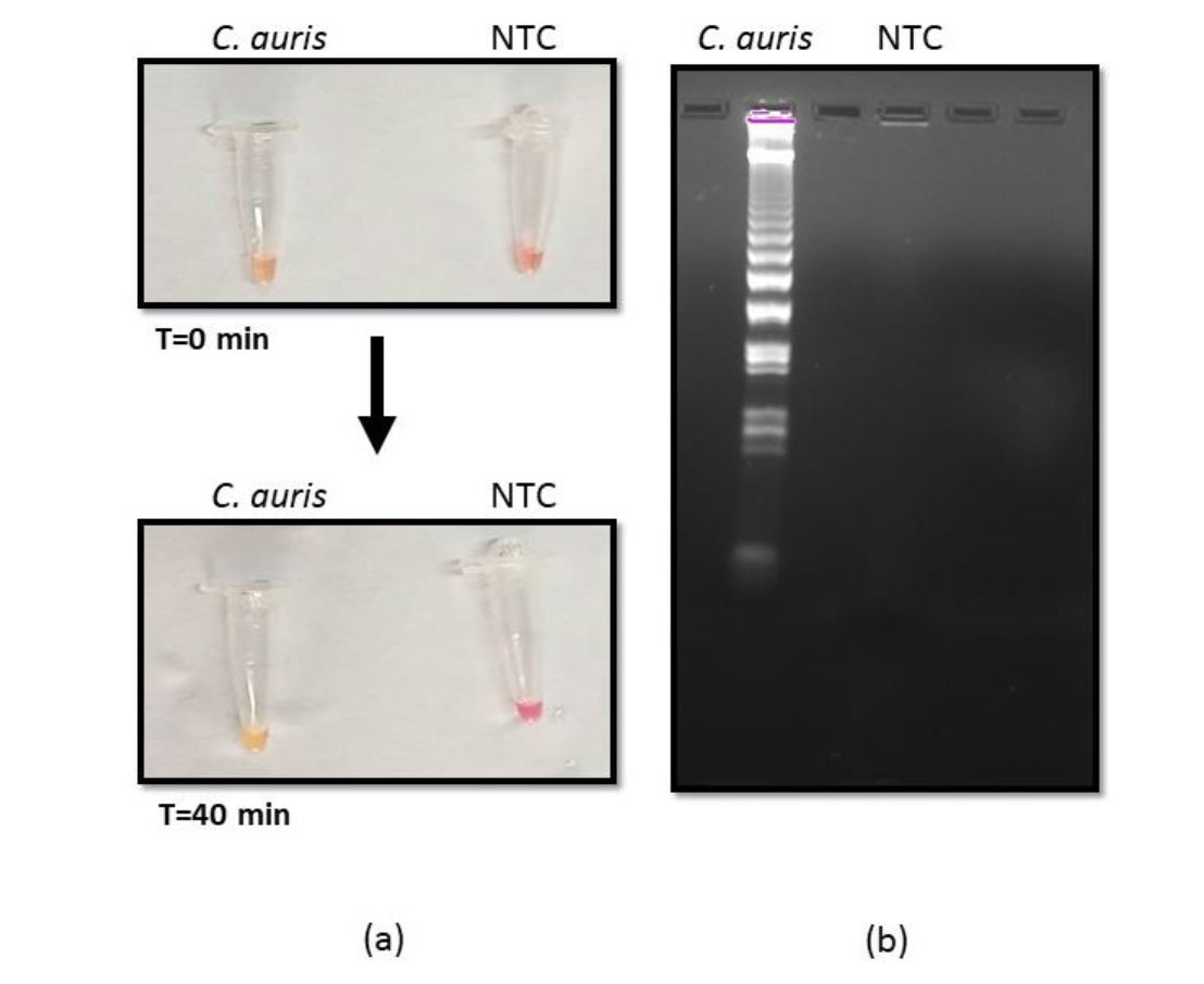
Figure 1. Optimization reverse transcription loop-mediated amplification (RT-LAMP) (a) RT-LAMP reaction with Candida auris RNA and negative template control visualized by color change from pink to yellow after 40 min at 65 ℃. (b) Agarose gel image of the amplified product obtained from LAMP reaction.
Analytical sensitivity of the RT-LAMP reaction
Different dilutions of extracted RNA from pure cultures of C. auris were used to determine the limit of detection (LOD) for our assay. The LOD was defined as the lowest dilution of RNA that was detectable by our assay with the naked eye. Firstly, the serial dilutions were tested with RNA concentrations ranging from 1,000 ng to 1 pg. It was found that the color change was noticeable at 1pg RNA concentration (Figure 2a; left panel). Furthermore, the researchers diluted the RNA until 1 ag concentration and again observed that color change was detectable even at 1 ag concentration (Figure 2a; right panel). Therefore, the LOD of the developed assay that was clearly visible both with the naked eye and gel electrophoresis (Figure 2b) was 1 ag of RNA.
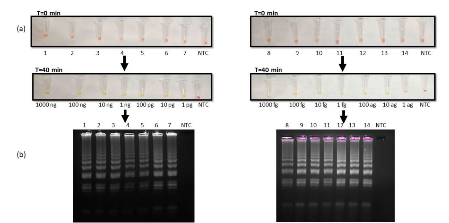
Figure 2. Limit of detection of reverse transcription loop-mediated amplification (RT-LAMP) reaction (a) RT-LAMP reaction in tubes 1-14 with RNA concentrations: Lane 1)1000 ng, 2)100 ng, 3)10 ng, 4)1ng, 5)100 pg, 6)10 pg, 7)1 pg (left panel) 8)1000 fg, 9)100 fg, 10)10 fg, 11)1 fg, 12)100 ag, 13)10 ag, 14)1 ag (right panel), visualised by color change from pink to yellow after 40 min at 65 ℃. (b) Agarose gel images of amplified products obtained from RT-LAMP reactions.
Additionally, this study sought to compare the analytical sensitivity of our newly developed RT-LAMP assay with the LAMP reaction using DNA as a template. This was achieved using different dilutions of extracted DNA from pure cultures of C. auris. Firstly, the serial dilutions were tested with DNA concentrations ranging from 1,000 pg to 10 fg. It was found that the reaction was sensitive at 10fg DNA concentration (Figure 3a; left panel). Furthermore, the researchers diluted the DNA until 1 ag concentration and observed that color change was still detectable only at 10 fg concentration (Figure 3a; right panel). These observations were also validated on gel electrophoresis (Figure 3b). Therefore, the LOD of the LAMP assay that was visible both with the naked eye and gel electrophoresis was 10 fg of DNA.
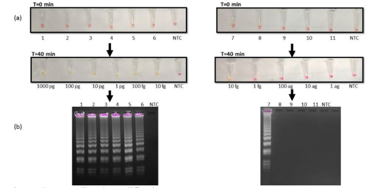
Figure 3. Lane 1)1000 pg, 2)100 pg, 3)10 pg, 4)1pg, 5)100 fg, 6)10 fg, (left panel), 7)10 fg, 8)1 fg, 9)100 ag, 10)10 ag, 11) 1 ag (right panel), visualized by colour change from pink to yellow after 40 minutes at 65℃. (b) Agarose gel images of amplified products obtained from LAMP reactions.
Specificity of RT- LAMP assay for C. auris detection
To evaluate the specificity of the assay in this study, different strains were used, viz., Mycobacterium smegmatis, C. albicans, and Escherichia coli other than C. auris. The RT-LAMP reaction was performed at 65 ºC for 40 min using template RNA isolated from Mycobacterium marinum, M. smegmatis, C. auris, and E. coli cultures. It was found that there was a clear amplification of only C. auris target PFOR gene as detected by naked eye owing to color change from pink to yellow contrary to retention of pink color in M. smegmatis, C. albicans, E. coli, and NTC (Figure 4a). This result was validated by gel electrophoresis of the amplicons depicting typical smear pattern only for C. auris (Figure 4b). Furthermore, intra-species specificity was also evaluated for the developed assay. Related Candida strains of C. auris, C. albicans, C. glabrata, C. tropicalis, C. parapsilosis, and C. krusei were included in the reaction. To our expectations, it was found that the amplification was achieved only with C. auris RNA which displayed yellow color contrary to all other Candida spp. RNA which retained their pink color (Figure 5a). Gel electrophoresis of the amplicons further validated the above-mentioned observations (Figure 5b).
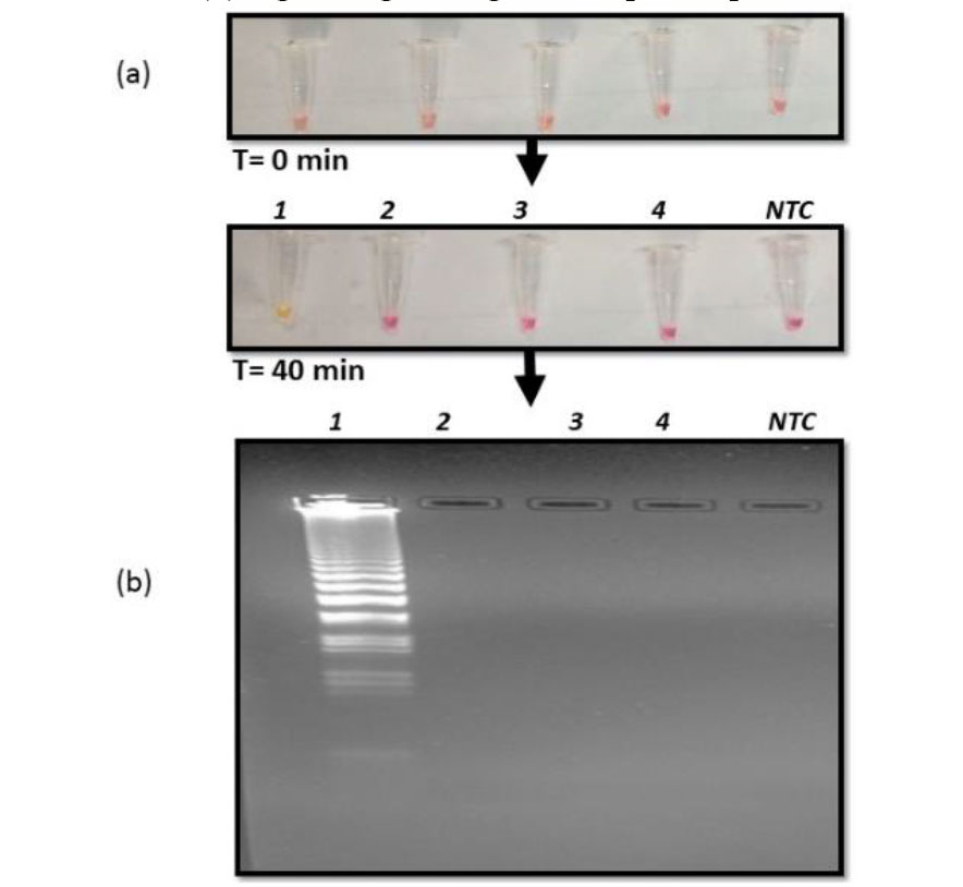
Figure 4. Interspecies specificity of reverse transcription loop-mediated amplification (RT-LAMP) reaction (a) RT-LAMP reaction of culture RNA extracted from Lane 1) C. auris, 2) C. albicans, 3) Mycobacterium smegmatis and 4) E coli, visualized by color change from pink to yellow after 40 minutes at 65℃. (b) Agarose gel image of the amplified product obtained from RT-LAMP reaction.
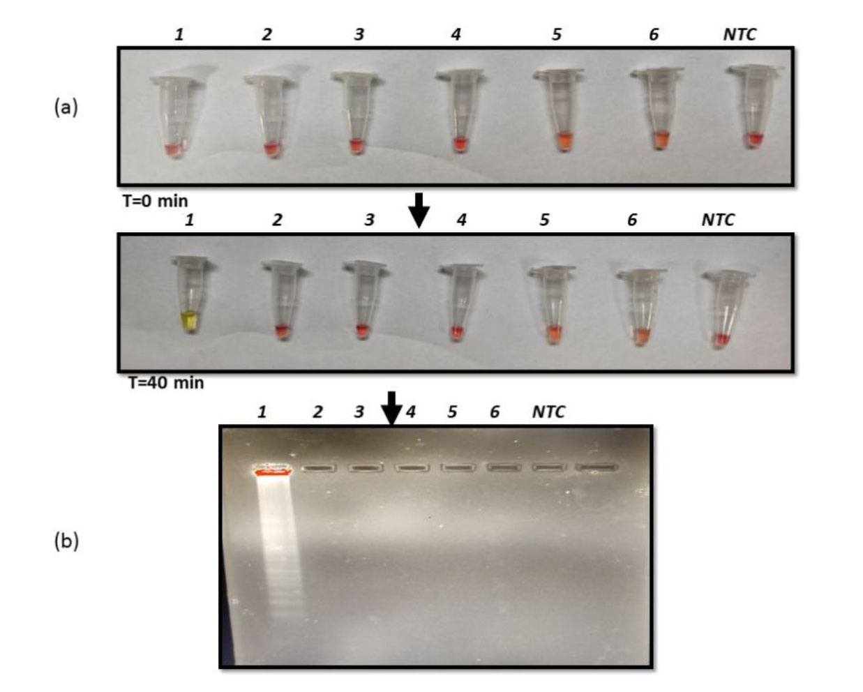
Figure 5. Intraspecies specificity reverse transcription loop-mediated amplification (RT-LAMP) reaction (a) RT-LAMP reaction of culture RNA extracted from lane 1) Candida auris, 2) Candida albicans, 3) Candida glabrata, 4) Candida tropicalis, 5) Candida parapsilosis, and 6) Candida krusei., visualized by color change from pink to yellow after 40 min at 65 °C. (b) Agarose gel image of the amplified product obtained from RT-LAMP reaction.
Clinical validation of RT- LAMP assay for C. auris detection
Validation of the developed assay was performed at the pilot level on 10 clinical isolates of C. auris. A clear amplification was found in all the tested C. auris strains by the naked eye owing to color change from pink to yellow contrary to the retention of pink color in NTC (Figure 6a). This result was again validated by gel electrophoresis of the amplicons depicting typical smear pattern for all tested isolates of C. auris (Figure 6b).
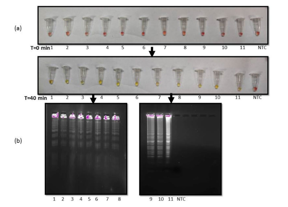
Figure 6. Clinical validation of reverse transcription loop-mediated amplification (RT-LAMP) for Candida auris (a) RT-LAMP reaction of culture RNA extracted from clinical isolates of C. auris in lanes 1) 1622, 2) 218834, 3) 1251, 4) C22/6089, 5) 37229, 6) 202916, 7) 202982, 8) P1188, 9) C-33, 10) C-34, 11) CBS10913T, and 12) NTC. (b) Agarose gel image of the amplified product obtained from RT-LAMP reaction.
Discussion
The rise in fungal infections among susceptible populations has brought C. auris as a rising obstacle against efficient therapeutics. In 2022, WHO declared C. auris a critical pathogen due to the complexities in its diagnosis leading to misidentification and treatment delays [ 1 ]. In contrast to laboratory testing, point-of-care testing offers a faster turnaround resulting in better clinical and economic outcomes as it overcomes the time-consuming steps of sample collection, transport, and processing by bringing the laboratory close to the patient. Rapid and accurate diagnosis with LAMP has emerged as a promising cost-effective method to detect Candida species due to its capability to detect visually, rapidly, and specifically [ 13 , 19 , 20 ]. The LAMP-based methods are much more sensitive and specific in comparison to culture and matrix-assisted laser desorption ionization-time of flight-based methods available in clinics for the diagnosis of C. albicans [ 21 ]. LAMP method was also able to detect vulvovaginal candidiasis with high sensitivity in a recent study [ 22 ]. Another study utilized the LAMP method to detect Candida with a sensitivity of 100% by using the blood sample from a patient [ 20 ]. The LAMP method was also used to detect C. glabrata with much sensitivity as compared to the culture method [ 23 ]. The novel LAMP diagnosis method was used for specific diagnosis of fungal species, such as Aspergillus fumigatus [ 24 , 25 ].
In recent years, a few variants of LAMP have also emerged which include RT-LAMP and multiplex LAMP (M-LAMP) [ 26 ]. The RT-LAMP method has been utilized for the detection of SARS-CoV-2 and is considered equivalent to RT-PCR in terms of utility [ 12 , 27 ]. Similarly, RT-LAMP has been used for the detection of parasites, such as amoeba and malaria [ 28 , 29 ]. In fact, the versatility of RT-LAMP has recently been reviewed for the early detection of pathogens [ 30 ]. Hence, the current study aimed to utilize the RT-LAMP approach to demonstrate its higher sensitivity and specificity, thereby, allowing faster detection and discrimination of C. auris from other microorganisms.
The present study used the PFOR domain of the C. auris genome that shared limited similarity with other Candida species based on the literature [ 12 ]. The specific primers were designed (Table 1) as described in the methods section and the RT-LAMP reaction was optimized at the temperature of 65 ºC for 40 min using RNA as template. The color change was detected by the naked eye and the typical gel electrophoresis pattern confirmed the successful amplification (Figure 1). Next, it was sought to determine the LOD by estimating the analytical sensitivity of the RT-LAMP assay. It was found that our assay was sensitive enough to give positive results till 1ag of RNA (Figure 2). Furthermore, the RT-LAMP was compared with the DNA LAMP method, and it was observed that unlike RNA, when DNA was used as a template, the LOD remained 10 fg (Figure 3). This confirmed that RT-LAMP was more sensitive than LAMP. This could also be attributed to the fact that RNA is abundant in copy numbers, including the splicing variants, compared to DNA; hence, it enables detection from metabolically active live cells contrary to dead cells. Another reason could be the single-chain RNA structure, which is more prone to degradation but less susceptible to contamination than DNA [ 17 , 31 ]. The Enhanced sensitivity of RT-LAMP can hold promise for rapid diagnosis of C. auris infections which are prone to misdiagnosis.
Furthermore, the specificity of the developed method was assessed using M. smegmatis, E. coli, and C. albicans strains. To our expectation, the RT-LAMP method could specifically discriminate C. auris from other tested strains (Figure 4). Additionally, this method was also deployed to test the intraspecies specificity and it was observed that color change could be detected only with C. auris RNA, contrary to other Candida strains (Figure 5). Based on these results, it was further sought to use the clinical isolates of C. auris to validate the assay. For this reason, 10 clinical isolates of C. auris were utilized. As it was expected, the researchers observed the color changes from pink to yellow for all tested clinical isolates of C. auris, contrary to NTC which remained pink (Figure 6a) as also apparent from gel electrophoresis (Figure 6b). This is also evident from the fact that LAMP-based methods are 10-100 times more sensitive, compared to the conventional PCR assays and other isothermal amplification techniques, such as CPA, PSR, or HAD [ 32 ]. The developed method in this study offers few advantages. For instance, the results can be interpreted with the naked eye owing to color change which makes the method suitable for limited-resource settings. Additionally, it is not dependent on equipment as it can be performed at a single temperature; therefore, these methods are cost-effective.
Conclusion
To win the battle against the disease, especially in resource-limited regions, simple, cost-effective, and rapid diagnostic methods are required for clinical application. In the era of isothermal-based amplification methods for disease diagnosis, the present study, although still in its infancy, offered considerable proof of concept for RT-LAMP-based methods that could be helpful in the rapid identification of C. auris infections. Development and improvement of these methods to meet the diagnostic gap in developing countries requires further investigation.
Acknowledgments
The authors are grateful to Sarman Singh and Arunaloke Chakrabarty for providing M. smegmatis mc2155 and C. auris strains as generous gifts, respectively. They would also like to thank the Central Instrumentation Research Facility, Amity University Haryana, for gel documentation experiments.
Authors’ contributions
A. K. was responsible for data curation, formal analysis, investigation, methodology, validation, visualization, and writing of the original draft. Z. F. contributed to conceptualization, project administration, resources, supervision, validation, and review and edition of the manuscript. S. H. played a key role in the conceptualization, formal analysis, investigation, methodology, project administration, resources, software development, supervision, validation, visualization, and writing of both the original draft and review and edition of the manuscript.
Conflicts of interest
No conflict of interest is declared.
Financial disclosure
This study received no specific funding.
References
- World Health Organization. WHO fungal priority pathogens list to guide research, development and public health action. World Health Organization. 2022.
- Satoh K, Makimura K, Hasumi Y, Nishiyama Y, Uchida K, Yamaguchi H. Candida auris sp. nov., a novel ascomycetous yeast isolated from the external ear canal of an inpatient in a Japanese hospital. Microbiol Immunol. 2009; 53(1):41-4.
- Cristina ML, Spagnolo AM, Sartini M, Carbone A, Oliva M, Schinca E, et al. An Overview on Candida auris in Healthcare Settings. J Fungi (Basel). 2023; 9(9):913.
- Sanyaolu A, Okorie C, Marinkovic A, Abbasi AF, Prakash S, Mangat J, et al. Candida auris: An Overview of the Emerging Drug-Resistant Fungal Infection. Infect Chemother. 2022; 54(2):236-46.
- Iguchi S, Itakura Y, Yoshida A, Kamada K, Mizushima R, Arai Y, et al. Candida auris: A pathogen difficult to identify, treat, and eradicate and its characteristics in Japanese strains. J Infect Chemother. 2019; 25(10):743-9.
- Borman AM, Fraser M, Johnson EM. CHROMagarTM Candida Plus: A novel chromogenic agar that permits the rapid identification of Candida auris. Med Mycol. 2021; 59(3):253-8.
- Khan Z, Ahmad S, Al-Sweih N, Joseph L, Alfouzan W, Asadzadeh M. Increasing prevalence, molecular characterization and antifungal drug susceptibility of serial Candida auris isolates in Kuwait. PLoS One. 2018; 13(4):e0195743.
- Ahmad S, Alfouzan W. Candida auris: Epidemiology, Diagnosis, Pathogenesis, Antifungal Susceptibility, and Infection Control Measures to Combat the Spread of Infections in Healthcare Facilities. Microorganisms. 2021; 9(4):807.
- Caceres DH, Forsberg K, Welsh RM, Sexton DJ, Lockhart SR, Jackson BR, et al. Candida auris: A Review of Recommendations for Detection and Control in Healthcare Settings. J Fungi (Basel). 2019; 5(4)
- Piepenburg O, Williams CH, Stemple DL, Armes NA. DNA detection using recombination proteins. PLoS Biol. 2006; 4(7):e204.
- Notomi T, Okayama H, Masubuchi H, Yonekawa T, Watanabe K, Amino N, et al. Loop-mediated isothermal amplification of DNA. Nucleic Acids Res. 2000; 28(12):E63.
- Bhatt A, Fatima Z, Ruwali M, Misra CS, Rangu SS, Rath D, et al. CLEVER assay: A visual and rapid RNA extraction-free detection of SARS-CoV-2 based on CRISPR-Cas integrated RT-LAMP technology. J Appl Microbiol. 2022; 133(2):410-21.
- Yamamoto M, Alshahni MM, Tamura T, Satoh K, Iguchi S, Kikuchi K, et al. Rapid Detection of Candida auris Based on Loop-Mediated Isothermal Amplification (LAMP). J Clin Microbiol. 2018; 56(9):e00591-18.
- Yadav R, Daroch P, Gupta P, Vaidya P, Mathew JL, Singh M, et al. Evaluation of TB-LAMP assay for detection of Mycobacterium tuberculosis in children. Infect Dis (Lond). 2021; 53(12):942-6.
- Morris U, Aydin-Schmidt B. Performance and Application of Commercially Available Loop-Mediated Isothermal Amplification (LAMP) Kits in Malaria Endemic and Non-Endemic Settings. Diagnostics (Basel). 2021; 11(2):336.
- Oscorbin I, Filipenko M. Bst polymerase - a humble relative of Taq polymerase. Comput Struct Biotechnol J. 2023; 21:4519-35.
- Wu D, Kang J, Li B, Sun D. Evaluation of the RT-LAMP and LAMP methods for detection of Mycobacterium tuberculosis. J Clin Lab Anal. 2018; 32(4):e22326.
- Ali B, Kumar M, Kumar P, Chauhan A, Usmani SA, Rudramurthy SM, et al. Sphingolipid diversity in Candida auris: unraveling interclade and drug resistance fingerprints. FEMS Yeast Res. 2024; 24:foae008.
- Bumbrah GS, Jain S, Singh S, Fatima Z, Hameed S. Diagnostic Efficacy of LAMP Assay for Human Fungal Pathogens: a Systematic Review and Meta-analysis. Curr Fungal Infect Rep. 2023;1-11.
- Wang Y, Zhao X, Zhou Y, Lu J, Yu H, Li S. Establishment and application of loop-mediated isothermal amplification coupled with nanoparticle-based lateral flow biosensor (LAMP-LFB) for visual and rapid diagnosis of Candida albicans in clinical samples. Front Bioeng Biotechnol. 2022; 10:1025083.
- Hernández Felices FJ, Tormo Palop N, Salvador García C, Mulet Bayona JV, Guna Serrano MR, Gimeno Cardona C. Evaluation of Eazyplex® LAMP test for fast Candida auris direct detection of colonized patients. Mycoses. 2024; 67(1):e13665.
- Li M, Jin X, Jiang Q, Wei H, Deng A, Mao Z, et al. Loop-Mediated Isothermal Amplification (LAMP): Potential Point-of-Care Testing for Vulvovaginal Candidiasis. J Fungi (Basel). 2023; 9(12):1159.
- Yahaya H, Cheah YK, Chee HY, Than LTL. Development and evaluation of a loop-mediated isothermal amplification (LAMP) method for Candida glabrata detection. Eur J Clin Microbiol Infect Dis. 2024 ; 43(11):2211-4.
- Owoicho O, Olwal CO, Tettevi EJ, Atu BO, Durugbo EU. Loop-mediated isothermal amplification for Candida species surveillance in under-resourced setting: a review of evidence. Expert Rev Mol Diagn. 2022; 22(6):643-53.
- Fallahi S, Babaei M, Rostami A, Mirahmadi H, Arab-Mazar Z, Sepahvand A. Diagnosis of Candida albicans: conventional diagnostic methods compared to the loop-mediated isothermal amplification (LAMP) assay. Arch Microbiol. 2020; 202(2): 275-82.
- Mori Y, Kitao M, Tomita N, Notomi T. Real-time turbidimetry of LAMP reaction for quantifying template DNA. J Biochem Biophys Methods. 2004; 59(2):145-57.
- Kitajima H, Tamura Y, Yoshida H, Kinoshita H, Katsuta H, Matsui C, et al. Clinical COVID-19 diagnostic methods: Comparison of reverse transcription loop-mediated isothermal amplification (RT-LAMP) and quantitative RT-PCR (qRT-PCR). J Clin Virol. 2021; 139:104813.
- Khunger S, Mewara A, Kaur U, Duseja A, Ray P, Kalra N, et al. Real-time loop-mediated isothermal amplification (real-time LAMP) assay for rapid diagnosis of amoebic liver abscess. Trop Med Int Health. 2024; 29(2):104-12.
- Buates S, Bantuchai S, Sattabongkot J, Han ET, Tsuboi T, Udomsangpetch R, et al. Development of a reverse transcription-loop-mediated isothermal amplification (RT-LAMP) for clinical detection of Plasmodium falciparum gametocytes. Parasitol Int. 2010; 59(3):414-20.
- Sen S, Bhowmik P, Tiwari S, Peleg Y, Bandyopadhyay B. Versatility of reverse transcriptase loop-mediated isothermal amplification (RT-LAMP) from diagnosis of early pathological infection to mutation detection in organisms. Mol Biol Rep. 2024; 51(1):211.
- Molaee N, Abtahi H, Ghannadzadeh MJ, Karimi M, Ghaznavi-Rad E. Application of Reverse Transcriptase -PCR (RT-PCR) for rapid detection of viable Escherichia coli in drinking water samples. J Environ Health Sci Eng. 2015; 13:24.
- Obande GA, Banga Singh KK. Current and Future Perspectives on Isothermal Nucleic Acid Amplification Technologies for Diagnosing Infections. Infect Drug Resist. 2020; 13:455-83.


