
Introduction
YEasts, including Candida species, are commensals on the skin, mucous membrane, gastrointestinal tract, and female genital tract. Candida albicans is considered the most common among clinically important Candida species. More than 50% of infections are caused by non-albicans Candida (NAC) species [ 1 ]. In recent years, an increasing number of invasive infections due to multidrug-resistant (MDR) Candida species have been reported worldwide. These emerging MDR Candida species include C. glabrata, C. krusei, C. guilliiermondii complex, C. rugosa, C. lusitaniae, C. lipolytica, C. haemulonii complex, C. kefyr, and C. auris. Among these potential MDR Candida species, C. auris has attracted more attention in recent years due to major outbreaks of invasive infections in healthcare facilities globally [ 1 , 2 ]. This is an emerging pathogen that is considered a threat to the global health system [ 3 ]. Following its first report in 2009 in Japan from a case of ear canal infection, C. auris has been reported worldwide including in India in various case reports, case series, and outbreaks in hospitals [ 4 - 9 ]. It has been found to be associated with various clinical conditions, including candidemia, urinary tract infections, pneumonia, and other invasive and non-invasive infections. Majority of the infections reported due to C. auris are among the patients with associated comorbidities or immunocompromised states. The patients admitted to intensive care units (ICU) or high-dependency units of the hospitals are at higher risk of getting infections with C. auris [ 5 - 9 ]. A resurgence of C. auris was reported among COVID-19 cases admitted to ICUs during the COVID-19 pandemic [ 10 , 11 ]. It is distinct from other Candida species due to its ability to acquire resistance to multiple antifungals and its propensity for transmission within a healthcare facility leading to outbreaks of hospital-acquired invasive infections associated with significant morbidity and mortality [ 12 , 13 ].
Reliable identification of C. auris using standard methods is difficult. The standard methods involve the panels of tests, including a combination of enzyme colorimetric tests, assimilation tests, and growth tests using carbohydrates and nitrogen compounds. Automated commercial identification systems rely upon disposable cards that contain sets of several conventional biochemical tests incorporated into microtiter wells used to identify a microorganism [ 14 ]. The reliable identification of C. auris can be carried out by matrix-assisted laser desorption ionization-time of flight mass spectrometry (MALDI-TOF MS) or various automated phenotypic identification systems, including VITEK-2 with updated database (version 8.01 or above) [ 14 , 15 ].
The data on C. auris infections is sparsely reported from India in case reports, case series, and a few outbreaks highlighting its clinical importance [ 4 - 9 ]. This study was planned to observe the occurrence of infections due to C. auris in a tertiary care medical teaching hospital in north India.
Materials and Methods
This cross-sectional hospital-based observational study was conducted in the Microbiology Department of a tertiary care medical teaching institute in north India for a period of 18 months from November 2022 to April 2024. The clinical specimens of all types were collected from the patients as per the standard specimen collection guidelines following inclusion and exclusion criteria. Patients of all age groups and genders showing the growth of only Candida species on culture were included in the study. The patients with prior administration of antifungals and growth of microorganisms other than Candida species were excluded from the study. The samples showing the growth of multiple isolates were also excluded from the study.
Institutional Ethics Committee Clearance
Approval of the institutional ethics committee was obtained before the commencement of the study with reference number SMC/UECM/2022/477B dated 14/10/2022.
Sample Processing
The clinical specimens were inoculated on Sabouraud dextrose agar (SDA) along with other conventional culture media for aerobic culture based on specimen type except blood culture. The blood culture was performed by BacT/ ALERT 3D automated blood culture system (bioMerieux, France) using respective blood culture bottles for pediatric and adult patients procured from bioMerieux, France. After flagging positive by the system, the broth from blood culture bottles was sub-cultured on SDA, blood agar, chocolate agar, and MacConkey agar. Any growth on solid culture media was further processed for presumptive identification by conventional microbiological procedures, including colony morphology and Gram staining. After preliminary identification of Candida, the species level identification was carried out by growth characteristics on chromogenic agar Candida (CHROMagar) and germ tube test. The culture media and reagents were procured from HiMedia India Private Limited, India. The final species level identification was performed by VITEK-2 compact automated system (bioMerieux, France) by using YST and YS408 cards for identification of Candida species and determination of minimum inhibitory concentration (MIC) for antifungal susceptibility testing (AFST), respectively, procured from bioMerieux, France. The quality control (QC) strains of Candida albicans ATCC 14053 and Candida parapsilosis ATCC 22019 were used for QC of VITEK-2 YST card and VITEK-2 AST YS408 cards, respectively, as recommended by bioMerieux. The colony characteristics of SDA having smooth and white-to-cream-colored colonies are shown in Figure 1. The microscopic findings of gram-stained smear depicting Gram-positive yeast-like cells are shown in Figure 2. Characteristics of C. albicans, C. tropicalis, C. parapsilosis, C. auris, C. kefyr, C. krusei, and C. dubliniensis on CHROMagar showing pigment production by various Candida species are illustrated in Figure 3.
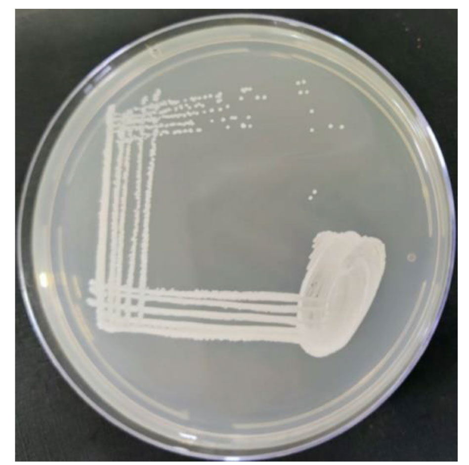
Figure 1. Colony characteristics of Candida auris on Sabouraud dextrose agar
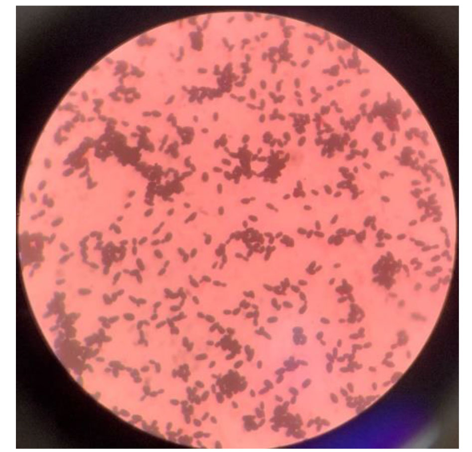
Figure 2. Microscopic features of Candida auris on Gram stain
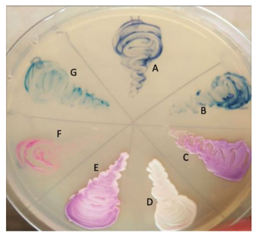
Figure 3. Characteristics of Candida auris on CHROMagar
As antifungal susceptibility breakpoints for C. auris are not established, hence the breakpoints for antifungal susceptibility of C. auris are defined based on the breakpoints established on expert opinion for closely related Candida species. The correlation between microbiologic antifungal breakpoints for C. auris and clinical outcome is not known. The MIC-based antifungal susceptibility breakpoints for C. auris as suggested by the Centers for Disease Control and Prevention (CDC) for AFST of C. auris were used in this study. The MIC-based antifungal susceptibility breakpoints as suggested by CDC for interpretation of AFST of C. auris are shown in Table 1 [ 16 ].
| Antifungal Name | Resistant Breakpoints (Minimum inhibitory concentration in µg/mL) |
|---|---|
| Fluconazole | ≥32 |
| Voriconazole | Based on susceptibility to fluconazole (surrogate marker for triazoles) |
| Amphotericin B | ≥2 |
| Caspofungin | ≥2 |
| Micafungin | ≥4 |
Statistical analysis
The data generated in this study was statistically analyzed in SPSS software (version 28.0, IBM Corp., Armonk, NY, USA). Categorical variables were compared using proportions and percentages.
Results
In total, 497 Candida isolates were isolated from the clinical samples collected during the study period. Out of the total Candida species, 106 (21.33%) were C. albicans while 391 (78.67%) were NAC species. Moreover, C. auris comprised 16 (3.22%) isolates of all Candida isolates and 4.09% of the NAC species.
Most of the C. auris isolates were recovered from adult patients (81.25%) followed by neonates (12.50%). Distribution of the demographic profile of the patients, including age, gender, and location, is summarized in Table 2.
| Parameter | Number (N=16) | |
|---|---|---|
| Age | ||
| Neonates (up to 1 month) | 2 (12.50%) | |
| Infants (1 month-1 year) | 0 | |
| Children (1-12 year) | 0 | |
| Adolescents (13-17 years) | 0 | |
| Adults (18-65 years) | 13 (81.25%) | |
| Elders (above 65 years) | 1 (6.25%) | |
| Gender | ||
| Male | 12 (75%) | |
| Female | 4 (25%) | |
| Location | ||
| Intensive care unit | 14 (87.50%) | |
| Surgical ward | 2 (12.50%) | |
Among various risk factors, ICU stay was the most common risk factor associated with C. auris infection comprising 14 (87.50%) cases. A summary of all the cases, including various risk factors, type of clinical specimen, and treatment outcome associated with C. auris infection, is tabulated in Table 3. The clinical conditions associated with C. auris infection were bloodstream infection and catheter-associated urinary tract infection comprising 50% each as shown in Table 3.
| Case No. | Age | Gender | Sample | Risk factors | Outcome |
|---|---|---|---|---|---|
| 1 | 5 days | Male | Blood | ICU Stay, presence of CVC, extreme of age | Survived |
| 2 | 6 days | Female | Blood | ICU Stay, presence of CVC, extreme of age | Deceased |
| 3 | 23 years | Male | Blood | ICU Stay, chronic kidney disease, steroid therapy | Survived |
| 4 | 24 years | Male | Blood | ICU Stay, steroid therapy | Survived |
| 5 | 29 years | Male | Urine | ICU Stay, presence of CVC, prior antibiotic exposure, indwelling urinary catheter | Survived |
| 6 | 29 years | Male | Blood | Surgery, prior antibiotic exposure, presence of CVC, indwelling urinary catheter, abdominal surgery | Survived |
| 7 | 30 years | Male | Blood | Surgery, prior antibiotic exposure, presence of CVC, indwelling urinary catheter, steroid therapy | Survived |
| 8 | 32 years | Male | Blood | ICU Stay, presence of CVC, indwelling urinary catheter, prior antibiotic exposure, steroid therapy | Survived |
| 9 | 34 years | Male | Urine | ICU Stay, indwelling urinary catheter | Survived |
| 10 | 49 years | Female | Urine | ICU Stay, indwelling urinary catheter, Diabetes mellitus | Survived |
| 11 | 53 years | Female | Urine | ICU Stay, indwelling urinary catheter, presence of CVC, steroid therapy, Cushing syndrome with morbid obesity | Survived |
| 12 | 54 years | Male | Urine | ICU Stay, indwelling urinary catheter, chronic kidney disease | Deceased |
| 13 | 62 years | Female | Urine | ICU Stay, indwelling urinary catheter, Diabetes mellitus | Survived |
| 14 | 76 years | Male | Urine | ICU Stay, indwelling urinary catheter, extreme of age, presence of CVC | Deceased |
| 15 | 51 years | Male | Urine | ICU Stay, indwelling urinary catheter, Diabetes mellitus | Survived |
| 16 | 41 years | Male | Blood | ICU Stay, presence of CVC, indwelling urinary catheter | Survived |
| ICU: Intensive care unit, CVC: Central venous catheter | |||||
The AFST data revealed the MDR nature of all C. auris isolates, which were found sensitive to echinocandins only. The antifungal susceptibility results of C. auris against fluconazole, voriconazole, caspofungin, micafungin, and amphotericin B are depicted in Figure 4.
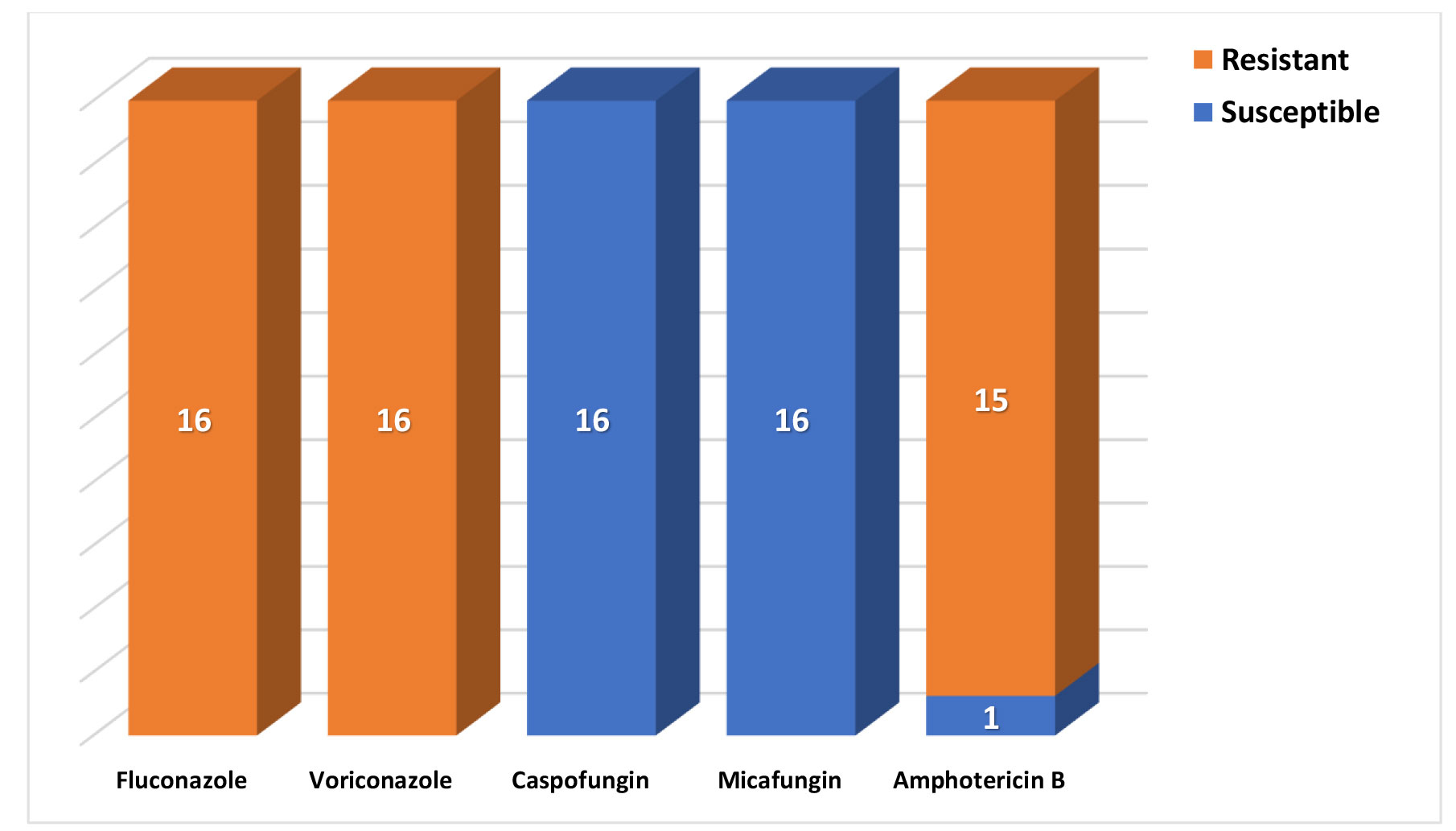
Figure 4. Antifungal susceptibility of Candida auris isolates
Discussion
After the first report on the isolation of C. auris in 2009 in Japan from a case of ear canal infection, it has been reported worldwide, including in India, in various case reports, case series, and outbreaks in hospitals [ 4 - 9 ]. In the present study, there was a 3.22% prevalence of C. auris among all Candida species isolated from clinical samples during the study period. Studies on the prevalence of C. auris are rarely available as most of the data on C. auris are either in the form of case reports, case series, or outbreaks in ICUs. The reported data on C. auris infections is only the tip of the iceberg as the actual data may be many folds since the identification of C. auris is a major issue in routine microbiology laboratories due to a lack of reliable identification infrastructure. The conventional methods are not reliable for identification and frequently lead to misidentification of C. auris as other biochemically related species of Candida. The reliable identification of C. auris can be performed by MALDI-TOF MS or various automated phenotypic identification systems, including VITEK-2 with an updated database (version 8.01 or above) [ 4 , 15 ].
The gender-wise distribution showed that the majority of the cases were males (75%), compared to females (25%). The higher isolation rates from males may be due to differences in the genetic and constitutional make of individual genders. Most of the case reports and case series also go in favor of the predominance of males. Cases of C. auris infection consisted of bloodstream infection and catheter-associated urinary tract infection each comprising 50% of the present study. The major proportion of cases were from ICUs (87.50%), compared to post-operative surgical wards (12.50%). Moreover, ICU stay was the single most common risk factor associated with C. auris infection. All of the cases in this study were associated with multiple risk factors and the common risk factors were ICU stay in 87.50%, indwelling urinary catheter in 75%, presence of CVC in 56.25%, and prior antibiotic exposure in 25% of the cases, while extreme of age, steroid therapy, surgical procedures, chronic kidney disease, and diabetes mellitus were among less common risk factors. Almost similar risk factors were also observed in other studies, including a review on the epidemiology of C. auris in healthcare facilities in Asia by Thatchanamoorthy et al. [ 17 ]. These stated various risk factors correlated well with the outbreaks from all over the Asian countries. Similar risk factors were also documented in other studies highlighting the opportunistic nature of C. auris [ 3 , 5 , 6 , 9 - 13 ].
A mortality rate of 18.75% due to C. auris was observed in the present study. There are variations in the reported data on mortality due to C. auris, as no mortality was reported from seven cases reported in an outbreak investigation in south India by Sathyapalan et al. [ 10 ]. Higher rates of mortality due to C. auris were also reported, including the studies performed by Ortiz-Roa et al. and Kaki in 2023 comprising mortality rates of 38.1% and 66.7%, respectively [ 18 , 19 ]. The mortality due to C. auris infections is higher, compared to that due to other Candida species [ 18 ]. The mortality and morbidity depend on multiple factors, such as delayed diagnosis leading to delayed initiation of targeted antifungal treatment, immunocompromised states, and associated comorbidities. The MDR nature of C. auris in the scenario of misidentification as other Candida species also contributes to the higher morbidity and mortality due to treatment failure. The antifungal susceptibility data highlighted the resistance of C. auris against most of the antifungal drugs and its susceptibility only to echinocandins, such as caspofungin and micafungin. Only one isolate of C. auris showed susceptibility against amphotericin B.
The limited available studies have highlighted the antifungal susceptibility data of C. auris. Almost similar findings were also observed in the studies performed by Prayag et al., Ashraf et al., and Sridharan et al. [ 20 - 22 ]. Since fluconazole is the most frequently used antifungal as an empirical medication in Indian hospitals, particularly in cases of unexplained fever or sepsis when the patient is not responding to antibiotics, this pattern of antifungal resistance is particularly concerning and disturbing. The echinocandins are advised as first-line antifungal treatment, subject to AFST due to the concern of azole and amphotericin B resistance in C. auris [ 21 , 23 ]. Reliable and accurate identification along with the AFST of C. auris is important for guiding therapy and evaluation of prognosis in ICUs, where MDR strains of the yeast are frequently prevalent [ 21 ]. If appropriate antifungal therapy is not timely instituted in conjunction with other interventions, such as the removal of invasive colonizing devices; invasive infections due to C. auris will likely result in a higher fatality rate [ 24 ].
Healthcare personnel in acute care settings have to use standard contact precautions to decrease the risk of transmission. Due to the MDR nature of C. auris isolates, as well as their ease of transmissibility in healthcare settings, it is imperative to quickly identify nosocomial outbreaks for the timely implementation of control measures. Since C. auris can live on a variety of surfaces, appropriate cleaning and disinfection techniques may be able to lower the risk of transmission [ 21 , 25 ]. As hand hygiene is the basic component of infection control, healthcare personnel should follow standard hand hygiene precautions to control the spread. The alcohol-based hand rubs are effective, as are chlorhexidine hand rubs against C. auris when hands are not visibly soiled. The contact precautions with gloves and gowns should be observed but gloves are not a substitute for hand hygiene [ 26 - 28 ].
There were certain limitations in this study. As this study was limited to a single hospital catering a a small proportion of the population, the findings cannot be generalized to the entire population. Moreover, molecular characterization of identification, including sequencing and genetic basis of antifungal drug resistance, was not studied due to limited resources.
Conclusion
Infections due to C. auris are emerging in hospital environments, especially among individuals having various risk factors, such as ICU stay, presence of indwelling devices, and immunocompromised states. The MDR nature of C. auris further complicates the situation as empirical antifungal therapy frequently results in treatment failure leading to higher mortality rates. Scarcity of reliable identification and AFST facilities are among other hurdles as conventional methods usually result in misidentification. Timely institution of appropriate antifungal treatment based on reliable identification and CDC-recommended antifungal susceptibility breakpoints is crucial to decrease mortality.
Acknowledgments
None.
Authors’ contributions
P. S. contributed to the concept, design, definition of intellectual content, literature search, data acquisition, data analysis, and manuscript preparation. A. P. contributed to the concept, design, and definition of intellectual content, manuscript preparation, manuscript edition, and manuscript review.
Conflicts of interest
None declared.
Financial disclosure
None.
References
- Ahmad S, Alfouzan W. Candida auris: Epidemiology, diagnosis, pathogenesis, antifungal susceptibility and infection control measures to combat the spread of infections in healthcare facilities. Microorganisms. 2021; 9(4):807.
- Colombo AL, Junior JNA, Guinea J. Emerging multidrug-resistant Candida species. Curr Opin Infect Dis. 2017; 30(6):528-38.
- Levy y, Miltgen G, Rousseau A, Lugagne N, Teysseyre L, Traversier N, et al. Case report: Emergence of Candida auris in the Indian Ocean Region. Am J Trop Med Hyg. 2021; 104(2):739-43.
- Satoh K, Makimura K, Hasumi Y, Nishiyama Y, Uchida K, Yamaguchi H. Candida auris sp. nov., a novel ascomycetous yeast isolated from the external ear canal of an inpatient in a Japanese hospital. Microbiol Immunol. 2009; 53(1):41-4.
- Rudramurthy SM, Chakrabarti A, Paul RA, Sood P, Kaur H, Capoor MR, et al. Candida auris candidaemia in Indian ICUs: analysis of risk factors. J Antimicrob Chemother. 2017; 72(6): 1794-801.
- Meena S, Rohilla R, Kaistha N, Singh A, Gupta P. Candida auris emergence in the Himalayan foothills: First case report from Uttarakhand, India. Current Medical Mycology. 2020; 6(1):47-50.
- Griffith N, Danziger L. Candida auris Urinary Tract Infections and Possible Treatment. Antibiotics (Basel). 2020; 9(12):898.
- Garcia Rivera MV, Heyl JJ, Oh MC. Candida auris Urinary Tract Infection in a nursing home patient with multicomorbities. Cureus. 2020; 12(12):e12322.
- Shaukat A, Ansari NA, Wali WA, Karic E, Madhoun IE, Mitwally H, et al. Experience of treating Candida auris cases at a general hospital in the state of Qatar. IDCases. 2020; 23:e01007.
- Sathyapalan DT, Antony R, Nampoothiri V, Kumar A, Shashindran N, James J, et al. Evaluating the measures taken to contain a Candida auris outbreak in a tertiary care hospital in South India: an outbreak investigational study. BMC Infectious Diseases. 2021; 21(1):425.
- Chowdhary A, Tarai B, Singh A, Sharma A. Multidrug-Resistant Candida auris Infections in Critically Ill Coronavirus Disease Patients, India, April–July 2020. Emerging Infectious Diseases. 2020; 26(11):2694-6.
- Calvo B, Melo AS, Perozo-Mena A, Hernandez M, Francisco EC, Hagen F, et al. First report of Candida auris in America: clinical and microbiological aspects of 18 episodes of candidemia. J Infect. 2016; 73(4):369-74.
- Morales-Lopez SE, Parra-Giraldo CM, Ceballos-Garzón A, Martínez HP, Rodríguez GJ, Álvarez-Moreno CA, et al. Invasive infections with multidrug-resistant yeast Candida auris, Colombia. Emerg Infect Dis. 2017; 23(1):162-4.
- Ambaraghassi G, Dufresne PJ, Dufresne SF, Vallières É, Muñoz JF, Cuomo CA, et al. Identification of Candida auris by use of the updated Vitek 2 yeast identification system, version 8.01: a multilaboratory evaluation study. J Clin Microbiol. 2019; 57(11): e00884-19.
- National Center for Emerging and Zoonotic Infectious Diseases. Centers for disease control and prevention. Algorithm to identify Candida auris based on phenotypic laboratory method and initial species identification. 2024.
- Centers for disease control and prevention. Antifungal Susceptibility Testing for C. auris. April 23, 2024.
- Thatchanamoorthy N, Devi VR, Chandramathi S, Tay ST. Candida auris: A mini review on epidemiology in healthcare facilities in Asia. J Fungi (Basel). 2022; 8(11):1126.
- Ortiz-Roa C, Valderrama-Rios MC, Sierra-Umana SF, Rodriguez JY, Muneton-Lopez GA, Solorzano-Ramos CA, et al. Mortality Caused by Candida auris Bloodstream Infections in Comparison with Other Candida Species, a Multicentre Retrospective Cohort. J Fungi (Basel). 2023; 9(7):715.
- Kaki R. Risk factors and mortality of the newly emerging Candida auris in a university hospital in Saudi Arabia. Mycology. 2023; 14(3):256-63.
- Prayag PS, Patwardhan S, Panchakshari S, Rajhans PA, Prayag A. The Dominance of Candida auris: A Single-center Experience of 79 Episodes of Candidemia from Western India. Indian J Crit Care Med. 2022; 26(5):560-3.
- Ashraf AA, Pinto R S, Karnaker VK. Candida auris in a Tertiary Healthcare Setting in South India: A Case Series. Curr Med Mycol. 2023; 9(4):33-8.
- Sridharan S, Gopalakrishnan R, Nambi PS, Kumar S, Nandini S, Ramasubramanian V. Clinical Profile of Non-neutropenic Patients with Invasive Candidiasis: A Retrospective Study in a Tertiary Care Center. Indian J Crit Care Med. 2021; 25(3):267-72.
- Kathuria S, Singh PK, Sharma C, Prakash A, Masih A, Kumar A, et al. Multidrug-resistant Candida auris misidentified as Candida haemulonii: characterization by matrix assisted laser desorption ionization–time of flight mass spectrometry and DNA sequencing and its antifungal susceptibility profile variability by Vitek 2, CLSI broth microdilution, and Etest method. J Clin Microbiol. 2015; 53(6):1823-30.
- de Araujo Motta F, Dalla-Costa LM, Dominguez Muro M, Lenzi A, Picharski GL, Burger M. Risk Adjustment for Congenital Heart Surgery Score as a Risk Factor for Candidemia in Children Undergoing Congenital Heart Defect Surgery. Pediatr Infect Dis J. 2016; 35(11):1194-8.
- Alvarado-Socarras JL, Vargas-Soler JA, Franco-Paredes C, Villegas-Lamus KC, Rojas-Torres JP, Rodriguez-Morales AJ. A Cluster of Neonatal Infections Caused by Candida auris at a Large Referral Center in Colombia. J Pediatric Infect Dis Soc. 2021; 10(5):549-55.
- Biswal M, Rudramurthy SM, Jain N, Shamanth AS, Sharma D, Jain K, et al. Controlling a possible outbreak of Candida auris infection: lessons learnt from multiple interventions. J Hosp Infect. 2017; 97(4):363-70.
- Abdolrasouli A, Armstrong-James D, Ryan L, Schelenz S. In vitro efficacy of disinfectants utilised for skin decolonisation and environmental decontamination during a hospital outbreak with Candida auris. Mycoses. 2017; 60(11):758-63.
- Sikora A, Hashmi MF, Zahra F. Candida auris. StatPearls Publishing. 2024.


