
Introduction
Candida species are frequent opportunistic and nosocomial pathogens in immunocompromised patients [ 1 ], associated with high mortality rates and prolonged hospital stay [ 2 ]. Worldwide studies have reported non-albicans Candida (NAC) species, such as C. glabrata, C. tropicalis, C. guilliermondii, C. krusei, C. lusitaniae, and C. parapsilosis as emerging pathogens, which are resistant to multiple antifungal agents [ 3 - 6 ]. Moreover, C. krusei and C. glabrata have shown reduced susceptibility to fluconazole [ 8 ] while C. lusitaniae are often resistant to amphotericin B [ 7 ], and intrinsically reduced susceptibility to echinocandins is seen in C. parapsilosis and C. guilliermondii [ 8 ]. Based on previous research, C. auris, a multidrug-resistant, healthcare-associated pathogen with a high mortality rate has particularly been a cause of concern [ 9 ]. Prolonged use of antifungal medications for prophylaxis or treatment of recurrent fungal infections in immunocompromised patients is a major factor leading to the emergence of antifungal resistance [ 10 ]. The epidemiological changes in the prevalence of the different Candida species causing systemic infections have been in parallel to the emergence of drug-resistant species [ 11 ]. In patients who received antifungal prophylaxis, NAC species were more commonly isolated than C. albicans [ 12 ]. During the COVID-19 pandemic, there was an increase in invasive fungal infections and subsequently the use of antifungal agents. Therefore, there have been reports of an increase in resistance to antifungal medications in the years following the pandemic [ 13 ]. This has led to a pressing need for continuous surveillance at the regional level to guide treatment. With this aim, the present study retrospectively analyzed the species distribution of Candida isolates and the pattern of antifungal drug susceptibility from various clinical samples at an urban tertiary care hospital in Eastern India, over a period of 5 years (2019- 2023).
Materials and Methods
This study received ethical approval from the institutional Ethical Committee (Registration number: ECR/62/Inst/WB/2013/RR-19). Samples from in-patient and out-patient departments (IPD and OPD) of our hospital in eastern India were processed in the hospital laboratory. Urine, high vaginal swabs, and blood cultures constituted the majority of the samples. Respiratory samples were not included in the study as Candida species are common colonizers of the respiratory tract and their role as pathogens is controversial [ 14 ]. Candida in urine was only reported in clinically symptomatic cases with pyuria, yielding pure growth of Candida in cultures and confirmed by repeated cultures whenever possible. High vaginal swabs were received mainly from the Department of Obstetrics for symptomatic cases and any growth of Candida spp. in these samples was reported since vaginal candidiasis in pregnant women is a risk factor for bloodstream infections, particularly in low birth weight and premature infants. Any growth in blood cultures and other samples was clinically correlated with the patient history. All Candida spp. that were reported over the period of 5 years (Jan 1, 2019, to December 31, 2023) were included in the study.
Samples were cultured on Sabouraud dextrose agar (SDA, HiMedia, India). Blood samples were incubated in blood culture bottles by BactAlert3D (Biomerieux, France) automated culture system. Bottles giving positive alarm within 5 days were subcultured on SDA and blood agar plates (Biomeriuex, France). Any yeast isolates were identified using the Vitek2 Compact system (Biomerieux, France) via ID-YST panel for identification and AST-YS08 panel for antifungal susceptibility testing to Amphotericin B, Caspofungin, Flucytosine, Fluconazole, Micafungin, and Voriconazole. The Clinical and Laboratory Standards Institute (CLSI M60, 2nd Edition, June 2020) and European Committee on Antimicrobial Susceptibility Testing (EUCAST) (Version 10.0, February 2020) breakpoints were used for the interpretation of the Vitek minimum inhibitory concentration (MIC) data. For the C. auris isolates, MICs were determined by broth microdilution method and interpreted using Centre for Disease Control (CDC) breakpoints as CLSI and EUCAST breakpoints were not defined. Statistical analysis was performed in SPSS software. Pearson's chi-squared test, Kruskal Wallis test, and one-way ANOVA tests were used to determine the association between categorical variables. It should be mentioned that P-values of less than 0.05 were considered statistically significant.
Results and Discussion
In total, 520 Candida species cultured from clinical samples were included in this study. Types of samples from which Candida was grown consisted of urine (68%) followed by high vaginal swabs (23.5%), blood (7.5%), and others (pus, ear swab, nasal mass biopsy tissue, and nappy rash) (1%). Majority of the samples were collected from the IPD (56%), followed by the intensive care units (ICU), high dependency units (HDU) (35%), and OPDs (9%). Based on findings, 24 (61.5%) out of 39 blood culture isolates were from the pediatric age group (<12 years) and 30 (77%) were from patients in critical care units (ICU, neonatal ICU, and Pediatric ICU).
Candida albicans isolates vastly outnumbered all other species of Candida isolated (70%) followed by C. tropicalis (22.5%). Other isolated species were C. guilliermondii, C. ciferrii, C. parapsilosis, C. glabrata, C. auris, C. lusitaniae (~1% each), C. pelliculosa, C. spherica, and C. krusei (~0.5% each). Number of patients with Candida growth were 87, 61, 65,141, and 128 in 2019, 2020, 2021, 2022, and 2023, respectively. In 2019, only 3 species of Candida were identified from our hospital (C. albicans, C. tropicalis, and C. ciferrii) but the diversity increased over the years to 10 species in 2023 (with the addition of C. auris, C. ciferrii, C. guilliermondii, C. glabrata, C. lusitaniae, C. spherica, and C. pelliculosa) [Figure 1].
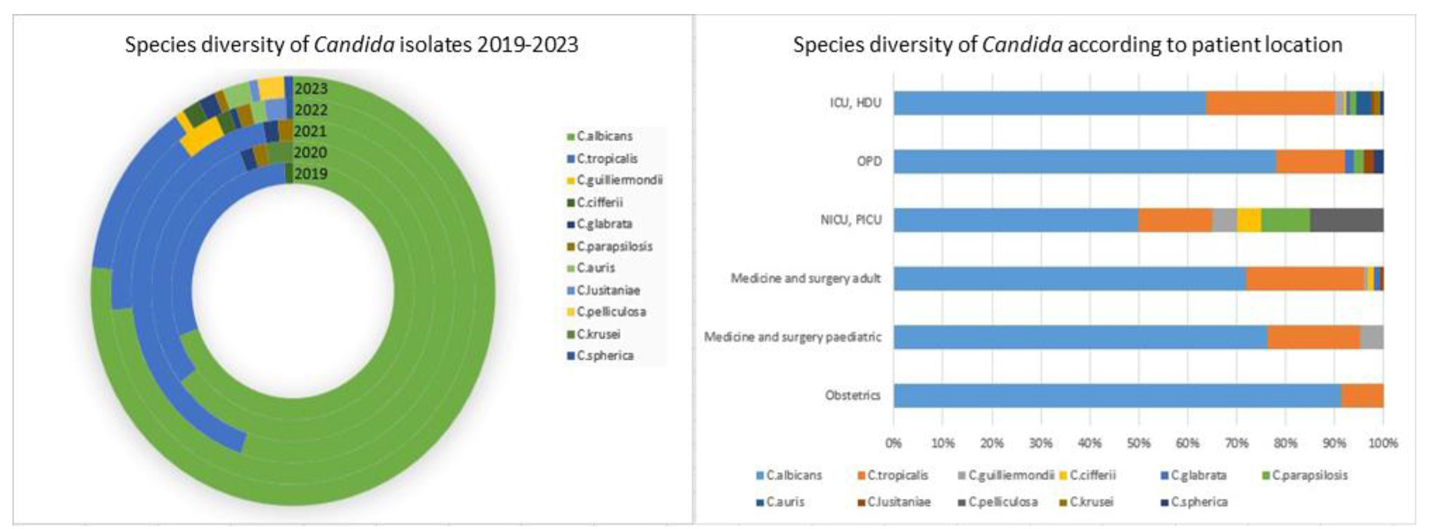
Figure 1. Species diversity of Candida isolated from clinical samples (2019-2023)
There were no C. auris isolated from 2019 to 2021, but between 2022 and 2023 there were five isolates of C. auris, all from patients in critical care units. One patient had C. auris isolated from both blood and urine denoting the likelihood of septicemia. However, the prevalence of C. auris in our center was much lower, compared to those reported in previous studies from India [ 15 - 17 ]. This could probably be due to the fact that these studies only considered samples from ICU setups. In terms of patient location, the least diverse samples were those collected from the obstetrics departments with only C. albicans and C. tropicalis being isolated while the most diverse samples, with a total of 10 species, were collected from the critical care departments (ICU and HDU) [Figure 1]. During the COVID-19 pandemic, patient intake and footfall were markedly reduced at the hospital due to unavailability of transportation, postponement of planned surgeries, and referral of COVID-19-positive cases to infectious disease facilities. However, it is evident that the number of cases of Candida infections remained quite high, and in the post-COVID period there was an increase in the number of cases which corroborated with reports of an increase in fungal infections as an aftermath of the COVID-19 pandemic [ 13 ].
Antifungal sensitivity patterns for C. albicans (365 isolates) and C. tropicalis (117 isolates) are summarized in Table 1.
| Name of drug | Candida albicans | Candida tropicalis | P- value | P-value | Candida albicans | Candida tropicalis | ||||||||||
|---|---|---|---|---|---|---|---|---|---|---|---|---|---|---|---|---|
| Total (5 years) | Total (5 years) | 2019 | 2020 | 2021 | 2022 | 2023 | P value | 2019 | 2020 | 2021 | 2022 | 2023 | P value | |||
| AMB | 16.71 | 10.26 | 0.090 | 0.281 | 1.64 | 16.67 | 2.70 | 23.28 | 22.94 | 0.0002 | 3.85 | 5.26 | 7.14 | 20 | 15.79 | 0.275 |
| CAS | 7.40 | 8.55 | 0.684 | 1.64 | 2.38 | 0 | 12.93 | 9.17 | 0.010 | 0 | 5.26 | 14.29 | 12.0 | 10.53 | 0.369 | |
| 5FC | 5.71 | 8.94 | 0.056 | 0 | 0 | 0 | 10.34 | 4.59 | 0.003 | 0 | 10.34 | 8.33 | 12.0 | 15.79 | 0.306 | |
| FLC | 5.21 | 6.84 | 0.504 | 0 | 2.38 | 2.70 | 11.21 | 3.67 | 0.008 | 0 | 15.79 | 7.14 | 8.00 | 5.26 | 0.359 | |
| MFG | 7.40 | 9.40 | 0.483 | 3.28 | 2.38 | 0 | 12.07 | 9.17 | 0.036 | 7.69 | 0 | 14.29 | 12.0 | 10.53 | 0.556 | |
| VRC | 13.70 | 5.13 | 0.011 | 6.56 | 11.9 | 8.11 | 17.24 | 16.51 | 0.225 | 0 | 5.26 | 7.14 | 8.00 | 5.26 | 0.733 | |
| AMB; Amphotericin B, CAS; Caspofungin, 5FC; Flucytosine, FLC; Fluconazole, MFG; Micafungin, Voriconazole; VRC | ||||||||||||||||
The other species were not included in this table due to their small numbers and the unavailability of complete CLSI and EUCAST breakpoints for many of them.
For C. albicans, the highest percentage of drug resistance over 5 years was observed against amphotericin B (17%) and voriconazole (14%), and in the case of C. tropicalis against amphotericin B (10%), caspofungin, flucytosine and micafungin (~9% each).
The MIC values for amphotericin B were also higher than those for the other antifungal agents. These findings differed from those of previous studies which found overall good sensitivity of Candida species to amphotericin B, and high resistance to the azoles, including fluconazole [ 18 - 25 ]. However, according to the CDC website, fluconazole resistance in Candida stands at around 7% which is similar to that observed in this study. There was no significant difference between C. albicans and C. tropicalis in terms of overall antifungal drug resistance (P=0.2819); however, when considering individual antifungal agents, C. albicans was significantly more resistant against voriconazole only, compared to C. tropicalis (P=0.0118). These findings are also in contrast to those of previous studies which described higher antifungal drug resistance in the NAC species, compared to C. albicans.
For C. albicans, the percentage of resistant isolates increased significantly over the period of 5 years (P<0.05) for all agents except voriconazole. For C. tropicalis, the increase in the percentage of resistant organisms was not statistically significant for any of the antifungal agents [Table 1].
Organisms with resistance to three or more antifungal drugs were considered multi-drug-resistant organisms (MDROs), and their total number was 31. Number of MDROs progressively increased over 5 years: there were no MDROs in 2019, 2 in 2020, 3 in 2021, 17 in 2022, and 9 in 2023. Majority of the MDROs were C. albicans (23), and the rest were C. tropicalis (8). Amongst the MDROs, the lowest resistance was observed against fluconazole with 12 of the 31 MDROs being susceptible to it.
Table 2 tabulates the MIC mode, MIC50, and MIC90 values of C. albicans and C. tropicalis each year. No significant change was noted in the mode of MIC values; however, there was a progressive increase in the MIC90 values, most markedly for amphotericin B for both organisms.
| Antifungal agent | Organism | MIC range (ug/ml) | Overall | 2019 | 2020 | 2021 | 2022 | 2023 | ||||||||||||
|---|---|---|---|---|---|---|---|---|---|---|---|---|---|---|---|---|---|---|---|---|
| Mode | MIC50 | MIC90 | Mode | MIC50 | MIC90 | Mode | MIC50 | MIC90 | Mode | MIC50 | MIC90 | Mode | MIC50 | MIC90 | Mode | MIC50 | MIC90 | |||
| AMB | C. albicans | ≤0.25 - ≥16 | 1 | 1 | 4 | 1 | 1 | 1 | 1 | 1 | 4 | 1 | 1 | 1 | 1 | 1 | 8 | 1 | 1 | 8 |
| C. tropicalis | ≤0.25 - ≥16 | 0.5 | 0.5 | 2 | ≤0.25 | 0.375 | 0.5 | ≤0.25 | ≤0.25 | 1 | ≤0.25 | 0.5 | 1 | ≤0.25 | 0.5 | 8 | ≤0.25 | 0.5 | ≥16 | |
| CAS | C. albicans | ≤0.12 - ≥8 | ≤0.12 | ≤0.12 | 0.25 | ≤0.12 | ≤0.12 | ≤0.12 | ≤0.12 | ≤0.12 | ≤0.12 | ≤0.12 | ≤0.12 | ≤0.12 | ≤0.12 | ≤0.12 | ≥8 | ≤0.12 | ≤0.12 | 0.25 |
| C. tropicalis | ≤0.12 - ≥8 | ≤0.12 | ≤0.12 | 0.5 | ≤0.12 | ≤0.12 | 0.25 | ≤0.12 | ≤0.12 | 0.25 | ≤0.12 | ≤0.12 | ≥8 | ≤0.12 | ≤0.12 | ≥8 | ≤0.12 | ≤0.12 | ≥8 | |
| 5FC | C. albicans | ≤1 - ≥64 | ≤1 | ≤1 | ≤1 | ≤1 | ≤1 | ≤1 | ≤1 | ≤1 | ≤1 | ≤1 | ≤1 | ≤1 | ≤1 | ≤1 | ≥64 | ≤1 | ≤1 | ≤1 |
| C. tropicalis | ≤1 - ≥64 | ≤1 | ≤1 | 16 | ≤1 | ≤1 | ≤1 | ≤1 | ≤1 | ≥64 | ≤1 | ≤1 | 16 | ≤1 | ≤1 | ≥64 | ≤1 | ≤0.5 | ≥64 | |
| FLC | C. albicans | ≤0.5 - ≥64 | ≤0.5 | ≤0.5 | 4 | ≤0.5 | ≤0.5 | 1 | ≤0.5 | ≤0.5 | 1 | ≤0.5 | ≤0.5 | 1 | ≤0.5 | ≤0.5 | 8 | ≤0.5 | ≤0.5 | 4 |
| C. tropicalis | ≤0.5 - ≥64 | ≤0.5 | 1 | 2 | ≤0.5 | ≤0.5 | 2 | 1 | 1 | 16 | 1 | 1 | 4 | ≤0.5 | ≤0.5 | 2 | ≤0.5 | ≤0.5 | 2 | |
| MFG | C. albicans | ≤0.06 - ≥8 | ≤0.06 | ≤0.06 | ≤0.06 | ≤0.06 | ≤0.06 | ≤0.06 | ≤0.06 | ≤0.06 | ≤0.06 | ≤0.06 | ≤0.06 | ≤0.06 | ≤0.06 | ≤0.06 | ≥8 | ≤0.06 | ≤0.06 | 0.12 |
| C. tropicalis | ≤0.06 - ≥8 | ≤0.06 | ≤0.06 | 0.5 | ≤0.06 | ≤0.06 | 0.5 | ≤0.06 | ≤0.06 | ≤0.06 | ≤0.06 | ≤0.06 | 4 | ≤0.06 | ≤0.06 | ≥8 | ≤0.06 | ≤0.06 | ≥8 | |
| VRC | C. albicans | ≤0.12 - ≥8 | ≤0.12 | ≤0.12 | 1 | ≤0.12 | ≤0.12 | ≤0.12 | ≤0.12 | ≤0.12 | 1 | ≤0.12 | ≤0.12 | ≤0.12 | ≤0.12 | ≤0.12 | 2 | ≤0.12 | ≤0.12 | 1 |
| C. tropicalis | ≤0.12 - ≥8 | ≤0.12 | ≤0.12 | ≤0.12 | ≤0.12 | ≤0.12 | ≤0.12 | ≤0.12 | ≤0.12 | ≤0.12 | ≤0.12 | ≤0.12 | ≤0.12 | ≤0.12 | ≤0.12 | ≤0.12 | ≤0.12 | ≤0.12 | ≤0.12 | |
| AMB; Amphotericin B, CAS; Caspofungin, 5FC; Flucytosine, FLC; Fluconazole, MFG; Micafungin, Voriconazole; VRC | ||||||||||||||||||||
Out of the five isolates of C. auris, all were susceptible to fluconazole, caspofungin, and micafungin, while two isolates were resistant to amphotericin B. The CDC breakpoints for C. auris against flucytosine and voriconazole were not available.
Therefore, an overall increasing trend of antifungal drug resistance as well as increasing diversity of species was noted in this 5-year period.
Conclusion
While antimicrobial drug resistance in bacteria is a major problem that is being addressed globally, fungal resistance to antifungal drugs is rarely highlighted and studied. In developing countries, such as India, empirical use of antifungal agents is common and has only increased following the rise of fungal infections during the COVID-19 pandemic. In this study, a considerable increase was found in the diversity of isolated species as well as the antifungal resistance over the period of 5 years in our hospital. Therefore, we should exercise caution in the use of antifungal drugs. Moreover, culture and sensitivity testing with robust clinical criteria for reporting fungal isolates should be followed in all suspected cases. Based on the findings of the present study, continuous surveillance of the prevalent species of Candida and their antifungal resistance patterns should be carried out in hospitals regularly, to guide treatment and prevent the spread of multidrug-resistant and nosocomial strains. A limitation of this study was its retrospective design which prevented the study of the mechanisms of drug resistance in these fungi, which is necessary to identify the means to overcome such resistance.
Acknowledgments
The authors would like to thank the laboratory staff of the Department of Microbiology, Ramakrishna Mission Seva Pratisthan for their contribution.
Authors’ contributions
P. M. compiled and analyzed all the data and prepared the manuscript. P. D. performed the statistical analysis. A. R. and P. M. reviewed the manuscript and provided valuable insights.
Conflicts of interest
None.
Financial disclosure
No additional funding was received for this study.
References
- Badiee P, Hashemizadeh A. Opportunistic invasive fungal infections: diagnosis & clinical management. Indian J Med Res. 2014; 139(2):195-204.
- Hirano R, Sakamoto Y, Kudo K, Ohnishi M. Retrospective analysis of mortality and Candida isolates of 75 patients with candidemia: a single hospital experience. Infect Drug Resist. 2015; 8:199-205.
- Chakrabarti A, Mohan B, Shrivastava SK, Marak RS, Ghosh A, Ray P. Change in distribution & antifungal susceptibility of Candida species isolated from Candidaemia cases in a tertiary care centre during 1996-2000. Ind J Med Res. 2002; 116:5-12.
- Ng KP, Saw TL, Na SL, Soo-Hoo TS. Systemic Candida infection in university hospital 1997-1999: the distribution of Candida biotypes and antifungal susceptibility patterns. Mycopathologia. 2001; 149(3):141-6.
- Mnge P, Okeleye BI, Vasaikar SD, Apalata T. Species distribution and antifungal susceptibility patterns of Candida isolates from a public tertiary teaching hospital in the Eastern Cape Province, South Africa. Braz J Med Biol Res. 2017; 50(6):e5797.
- Orasch C, Marchetti O, Garbino J, Schrenzel J, Zimmerli S, Mühlethaler K, et al. Candida species distribution and antifungal susceptibility testing according to European Committee on Antimicrobial Susceptibility Testing and new vs. old Clinical and Laboratory Standards Institute clinical breakpoints: a 6-year prospective Candidaemia survey from the fungal infection network of Switzerland. Clin Microbiol Infect. 2014; 20(7):698-705.
- Perea S, Patterson TF. Antifungal Resistance in Pathogenic Fungi. Clin Infect Dis. 2002; 35 (9):1073-80.
- Perlin DS. Echinocandin Resistance in Candida. Clin Infect Dis. 2015; 61(6):S612-7.
- Chowdhary A, Sharma C, Meis JF. Candida auris: A rapidly emerging cause of hospital-acquired multidrug-resistant fungal infections globally. PLoS Pathog. 2017; 13(5):e1006290.
- Sanglard D. Emerging Threats in Antifungal-Resistant Fungal Pathogens. Front Med. 2016; 3:11.
- Fournier P, Schwebel C, Maubon D, Vesin A, Lebeau B, Foroni L, et al. Antifungal use influences Candida species distribution and susceptibility in the intensive care unit. J Antimicrob Chemother. 2011; 66(12):2880-6.
- Bassetti M, Ansaldi F, Nicolini L, Malfatto E, Molinari MP, Mussap M, et al. Incidence of Candidaemia and relationship with fluconazole use in an intensive care unit. J Antimicrob Chemother. 2009; 64:625-9.
- Pfaller MA, Carvalhaes CG, DeVries S, Rhomberg PR, Castanheira M. Impact of COVID-19 on the antifungal susceptibility profiles of isolates collected in a global surveillance program that monitors invasive fungal infections. Med Mycol. 2022; 60(5):myac028.
- Pendleton KM, Huffnagle GB, Dickson RP. The significance of Candida in the human respiratory tract: our evolving understanding. Pathog Dis. 2017; 75(3):ftx029.
- Mathur P, Hasan F, Singh PK, Malhotra R, Walia K, Chowdhary A. Five-year profile of Candidaemia at an Indian trauma centre: High rates of Candida auris blood stream infections. Mycoses. 2018; 61(9):674-80.
- Rudramurthy SM, Chakrabarti A, Paul RA, Sood P, Kaur H, Capoor MR, et al. Candida auris Candidaemia in Indian ICUs: analysis of risk factors. J Antimicrob Chemother. 2017; 72(6):1794-801.
- Chowdhary A, Tarai B, Singh A, Sharma A. Multidrug-Resistant Candida auris Infections in Critically Ill Coronavirus Disease Patients, India, April-July 2020. Emerg Infect Dis. 2020; 26(11):2694-6.
- Bhattacharjee P. Epidemiology and antifungal susceptibility of Candida species in a tertiary care hospital, Kolkata, India. Curr Med Mycol. 2016; 2(2):20-7.
- Thomas M, Oberoi A, Dewan E. Species distribution and antifungal susceptibility of candidemia at a multispecialty center in North India. CHRISMED J Health Res. 2016; 3(1):33-6.
- Vaghela GM, Purohit LN, Kariya PV. Susceptibility of Candida species to antifungal drugs in Western India. Nat J Med Res. 2015; 5(2):122-6.
- Pahwa N, Kumar R, Nirkhiwale S, Bandi A. Species distribution and drug susceptibility of Candida in clinical isolates from a tertiary care centre at Indore. Ind J Med Microbiol. 2014; 32(1):44-8.
- Basu S, Gugnani HC, Joshi S, Gupta N. Distribution of Candida species in different clinical sources in Delhi, India, and proteinase and phospholipase activity of Candida albicans isolates. Rev Iberoam Micol. 2003; 20(4):137-40.
- Dutta V, Lyngdoh WV, Bora I, Choudhury B, Khyriem AB, Bhattacharyya P. Characterization of Candida species from Intensive Care Unit Isolates in a Tertiary Care Centre in North-East India: A retrospective study. Int J Med Public Health. 2015; 5(4):312-6.
- Kurup Rajgopal S. Prevalence and Antifungal Susceptibilities of Candida spp from a South Indian Tertiary Care Hospital. Int J Med Res and Rev. 2017; 5(2):98-104.
- Giri S, Kindo AJ, Kalyani J. Candidemia in intensive care unit patients: A one year study from a tertiary care center in South India. J Post Grad Med. 2013; 59(3):190-5.

