
Introduction
Candida species are among the most prevalent aetiologic agents in human invasive infections, with a reported mortality rate of 40-51% [ 1 , 2 ]. Immunocompromised patients and those hospitalized in intensive care units with horizontal transmission are at a higher risk of invasive candidiasis (IC) [ 3 - 5 ].
Although Candida albicans has been implicated in the majority of IC cases, the incidence rate of non-albicans Candida (NAC) species is rapidly rising, with Candida glabrata reported as the second most common cause of candidemia after C. albicans [ 6 - 8 ], with morbidity and mortality rates of 40-60%. [ 9 ].
Currently, only three classes of medications are approved for the treatment of systemic fungal infections [ 10 - 12 ]. Azoles have been widely used as the first-line treatment of IC [ 13 - 15 ], but several reports of azole-resistant strains of C. albicans and NAC species have emerged, and another major antifungal class, echinocandins, is now used as first-line therapy for the treatment of IC in many countries [ 15 , 16 ]. Most Candida species are susceptible to echinocandins and amphotericin B, although a high incidence rate of C. glabrata resistance against echinocandins has been reported [ 16 , 17 ], usually accompanied by cross-resistance to azoles [ 18 , 19 ] and reduced sensitivity to amphotericin B [ 20 ]. Medication resistance has been attributed to long-term exposure to antifungal medications [ 3 , 5 , 21 , 22 ] and biofilm formation [ 23 ].
Sessile cells (within the biofilm) differ from planktonic (free-living) populations [ 24 ] in showing higher antifungal resistance levels [ 25 ]. Candida biofilms are enclosed in an extracellular matrix (ECM) of carbohydrates (e.g., polysaccharides), proteins, and DNA [ 26 , 27 ]. The ECM of the Candida biofilm contributes to pathogenicity by increasing medication tolerance and promotion of immune evasion [ 28 - 30 ], making biofilm formation an important virulence factor of Candida species and a cause of treatment failure [ 31 , 32 ].
In its quest to focus on the increasing burden of antimicrobial resistance and to guide and promote research to improve diagnostic and treatment strategies, the World Health Organization categorized C. albicans and C. glabrata as critical and high-priority pathogens, respectively, in its first published fungal pathogen priority list [ 33 ]. Development of antifungal medication resistance in C. albicans and NAC species poses a serious challenge [ 34 ] and creates an urgent need for the identification and development of novel antifungal agents.
Antifungal compounds of plant origin are therapeutic alternatives that can potentially be used to curb the increasing number of fungal infections [ 35 - 37 ]. South Africa is home to the richest temperate flora in the world; 11,700 out of 19,581 indigenous plant species are endemic and 5,000 species are used as medicines, with 350 species commonly traded as medicinal plants [ 38 , 39 ]. Galenia africana, also known as “kraalbos”, “geelbos” or “perdebos”, belongs to the Aizoaceae family and is a local medicinal plant predominantly found in the Namaqualand and Karoo areas, extending to the Eastern Cape of South Africa [ 40 ]. Hydroethanolic extracts of G. africana have shown antifungal activity against 14 different fungal species in vitro [ 41 ], with greater antifungal activity and synergy with fluconazole demonstrated with ethanolic extracts against planktonic C. albicans and C. glabrata isolates [ 42 ].
Impedance sensors have several applications, including the evaluation of microbial biofilm formation [ 43 , 44 ]. The xCELLigence RTCA instrument (Cat. no. 05469759001, ACEA Biosciences, USA) can detect relative changes in conductivity caused by the interaction between cells adhered to the bottom of the test plate and gold microelectrodes in the presence of an electrically conductive solution [ 44 ], allowing for continuous monitoring of the adhesion properties of cells in a non-invasive label-free manner [ 45 ]. The electrical impedance is measured and recorded by the xCELLigence software as a cell index (CI), the magnitude of which is dependent on the cell number, cell morphology and size, and the strength of cell attachment to the surface of the plate [ 45 - 47 ]. The xCELLigence system has been used to measure the biofilm formation of C. albicans [ 48 , 49 ] and Candida interspecies biofilm interactions [ 50 ]. However, its application in testing the antifungal action of medicinal plants against Candida has not been reported.
Therefore, the present study aimed to use real-time impedance-based technology to investigate the ability of G. africana to disrupt existing C. albicans and C. glabrata biofilms and prevent biofilm formation.
Materials and Methods
Galenia africana extract preparation
Galenia africana was supplied as a 20% (w/v) air-dried extract of leaves and shoots, commercially prepared (Brenn-O-Ken Pty Ltd, Wolseley, South Africa) with 60% ethanol and oven-dried under negative pressure to produce crystals. Permission to conduct research on this indigenous plant was obtained from the South African Department of Forestry, Fisheries, and the Environment (Registration Number BABS 000314). A stock solution was prepared by weighing the dried crystal extract of G. africana and constituting it in a 50 mL centrifuge tube containing RPMI 1640 medium (Cat. no. R6504, Sigma-Aldrich, St. Louis, MI, USA). Sterile forceps were used to crush and dissolve the extract, with intermittent vortexing. The extract was incubated at approximately 20-25 °C for 24 h. This was followed by final crushing and vortexing to allow the phytochemicals of the extract to dissolve into the media. The tubes were centrifuged for 10 min at 20,000 rpm, and the supernatant was aseptically aspirated and transferred to a 15 mL centrifuge tube. Tubes were further centrifuged at 4,000 rpm for 5 min. This process was repeated several times until a debris-free, brown-green plant tincture was observed. The tincture was filter sterilized using a 5 mL sterile syringe and a 0.22 μm filter into a 2 mL sterile microcentrifuge tube. Stock concentrations (yielding a 400 mg/mL stock solution, equivalent to a 40% (w/v) extract concentration) were stored at 4 °C.
Candida species and culture conditions
Candida albicans (ATCC 90028) and Candida glabrata (ATCC 26512), obtained from the American Type Culture Collections (ATCC, Manassas, VA, USA) were used for the real-time analysis of adherence and subsequent biofilm formation. The isolates were revived by growth in 10 mL Sabouraud dextrose broth (Cat. no. CMO147, Oxoid, UK), followed by incubation at 37 °C for 3-5 days. Purity of growth was confirmed by microscopy and subculture of single colonies previously grown on Sabouraud dextrose agar (SDA) (Cat. no. 84088, Sigma-Aldrich, St. Louis, MI, USA) and incubated aerobically at 37 °C for 24-48 h. Species differentiation was confirmed by growth on Fluka chromogenic Candida identification agar (Cat. no. 94382; Sigma-Aldrich, St. Louis, MI, USA) and Oxoid chromogenic Candida agar (Cat. no. CM1002A; Oxoid, Hampshire, UK) at 30 °C for 24-72 h. The type strains were subcultured onto SDA plates for 24 h at 37 °C and individual colonies were picked to inoculate 7 mL of fresh, sterile yeast peptone dextrose (YPD) broth (Cat. no. Y1375, Sigma-Aldrich, St. Louis, MI, USA). A nephelometer (Cat. no. V3011, ThermoFisher Scientific, Waltham, MA, USA) was used to adjust the growth to a 0.5 McFarland standard suspension, yielding a cell suspension of approximately 1×10⁶-5×10⁶ CFU/mL. This inoculum was further diluted and optimized to a working suspension of 1:20 in YPD broth.
Real-time monitoring of Candida species adhesion and disruption of 10-h preformed biofilm formation
Real-time cell adhesion monitoring and subsequent biofilm assays were performed using the xCELLigence real-time cell analysis-dual purpose (RTCA-DP) instrument (Cat. no. 05469759001, ACEA Biosciences, USA). Due to the high cost of the xCELLigence gold electrode-embedded plates, experiments were performed in duplicate, with the test repeated in cases of discrepancy. The YPD broth optimized working suspension was used as the culture medium for the growth of the isolates in the E-plate 16 (Cat. no. 0546830001, ACEA Biosciences, USA), as previously described [ 49 ], since this medium is known to promote Candida adherence and biofilm formation [ 50 , 51 ]. The E-plates were prepared by adding fresh, sterile YPD broth (100 µL) to each well. The plates were left at 20-25 °C for 30 min to achieve equilibrium between the culture media and the E-plate surface. After equilibration of the instrument at 37 ºC and the E-plates at 20-25 °C, plates were inserted into the chamber of the RTCA-DP instrument, and the RTCA software package (Cat. No. 05454433001, ACEA Biosciences, USA) was used to measure the background impedance for each well. Candida suspensions (equivalent to 0.5 McFarland standard) were added to the wells of the E-plates containing 100 µL of YPD broth. Each well received 50 µL of the inoculum (as this volume provides less variation in seeding density). Sterility and growth control wells were included in each plate. All the wells contained a final volume of 150 µL/well, except the sterility control well, which contained a final volume of 200 µL of YPD. The inoculated E-plates were left at 20-25 °C for 30 min to allow the cells to settle to the bottom of the wells. Sterile distilled water was added to the surrounding evaporation-control troughs, as recommended by the manufacturer. The plates were placed into the cradle of the RTCA-DP instrument, allowing the Candida-type strains to enter the exponential growth phase, at approximately 10 h. An experimental procedure was logged on the RTCA software package, with impedance readings set to take place at 15-minute intervals for 10 h. Following the incubation step, the E-plates were removed from the instrument, followed by the addition of various concentrations of G. africana, with the final concentrations within the inoculated wells ranging from 0.78 mg/mL to 50 mg/mL. Subsequently, 50 µL of the plant extract was added to the designated wells, bringing the final volume to 200 µL. The plates were returned to the instrument and further incubated at 37 °C with impedance recorded every 15 min for an additional 38 h, bringing the final incubation time to 48 h.
Real-time monitoring of Candida spp. adhesion and prevention of initial biofilm formation
Ability of G. africana to prevent Candida species adhesion and subsequent biofilm formation was evaluated using impedance-based technology, employing the same preliminary and optimization steps. However, after the equilibration steps, the wells of the E-plates were inoculated with 50 µL of various concentrations of the G. africana extract, ranging from 0.78 mg/mL to 50 mg/mL, followed by the addition of 50 µL of the optimized fungal suspension. The control wells were treated as described above. The E-plates were placed into the cradle of the RTCA-DP instrument and incubated for a total of 48 h at 37 °C, with impedance measured every 15 min.
Results
Disruption of 10-hour preformed Candida albicans adhesion and subsequent biofilm formation
Prior to the determination of the critical time point at which the adhered fungal cells would be challenged with the extract (e.g., the window of treatment of preformed Candida species biofilms) [ 45 ], cellular behavior and optimized medication treatment times were observed. During the initial stage of the CI curves, the Candida species entered a biofilm formation exponential phase at approximately 7 h, followed by entry into the mid-exponential growth phase at approximately 10 h, where CI values increased, representing adhesion and subsequent biofilm formation. Candida biofilm formation increased exponentially up to 15 h. Therefore, G. africana crude extract was administered at approximately 10 h post-seeding of the Candida-type strains, at the start of the mid-exponential growth phase.
Figures 1 and 2 represent the mean CI curves obtained for the duplicate experiments of the same biological sample, following a 48-h total incubation period. The curves followed the same pattern for each repeated experiment, with no discrepancies observed. In the present study, a plateau phase was observed at approximately 20 h post-incubation. The CI profile revealed that among the two Candida spp. tested, C. albicans demonstrated a stronger adherence and subsequent biofilm formation, compared to C. glabrata. A comparison of CI values of the positive (untreated) controls of the two Candida species revealed that C. albicans had a maximum CI of 1.50 at approximately 15 h and C. glabrata had a maximum CI of 0.89 at approximately 10 h.
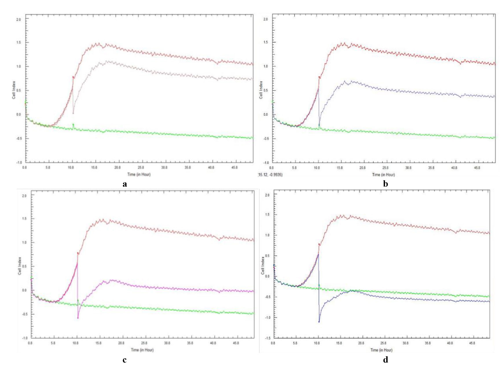
Figure 1. Real-time cell analysis cell index variations of 10-hour preformed Candida albicans (ATCC 90028) cell adhesion and subsequent biofilm formation treated with ascending concentrations of Galenia africana aqueous extract: a) 6.25 mg/mL, b) 12.5 mg/mL, c) 25 mg/mL, and d) 50 mg/mL. A positive control (burgundy curve) and sterility control (green curve) are included.
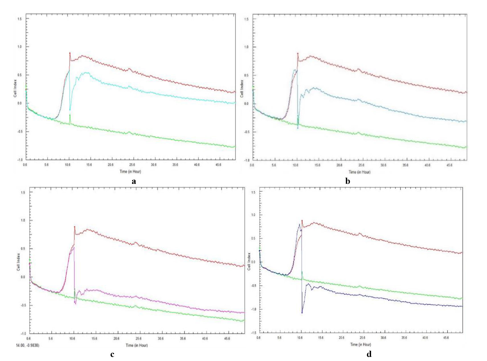
Figure 2. Real-time cell analysis cell index variations of 10-hour preformed Candida glabrata (ATCC 26512) cell adhesion and subsequent biofilm formation treated with ascending concentrations of Galenia africana aqueous extract: a) 6.25 mg/mL, b) 12.5 mg/mL, c) 25 mg/mL, and d) 50 mg/mL. A positive control (burgundy curve) and sterility control (green curve) are included.
When testing C. albicans with G. africana, the 6.25 mg/mL concentration (Figure 1a) resulted in a noticeable decrease in CI (0.06) after the addition of the extract at 10 h, compared to the positive control (0.7). This was observed by an immediate and transient decrease in the CI value. However, this concentration was incapable of completely eradicating or disrupting the adhesion and biofilm formation as the CI values remained positive at the end of the incubation period. Higher concentrations of G. africana, namely, 25 and 50 mg/mL (Figure 1c and d), proved to be the most effective in completely disrupting and removing the adhesion and biofilm formation. The CI values, -0.58 and -1.10, were the lowest values after the addition of G. africana extract at 10 h, in that order, compared to the CI of the growth control, with the 50 mg/mL concentration showing complete removal of the adhesion and biofilm formation. These results indicated that no adhesion and subsequent biofilm formation were present, with CI values of 0.0 and -0.5, at the end of the experiment.
As with C. albicans, the lower concentrations of G. africana did not appear to significantly disrupt the biofilm formation of C. glabrata, as adhesion was still evident with positive CI values, similar to the positive control at the end of the experiment. However, the 6.25 mg/mL G. africana concentration (Figure 2a) resulted in a noticeable decrease in CI value (-0.13) after the addition of the extract at 10 h, compared to the positive control (0.9). This was also observed in the other concentrations, with an immediate and transient decrease in the CI value. Similar to the results obtained for C. albicans, the lowest concentration was incapable of completely eradicating or disrupting the adhesion and biofilm formation, as the CI values remained positive at the end of incubation, while the higher concentrations of G. africana tested, namely 25 and 50 mg/mL (Figure 2c and d), proved to be the most effective in completely disrupting and removing the adhesion and biofilm formation.
Prevention of Candida species adhesion and biofilm formation
Figures 3 and 4 represent the CI curves following a 48-h total incubation period. The curves followed the same pattern for each repeated experiment, with no discrepancies observed. The real-time curves relate to the ability of each concentration of G. africana to prevent the initial adhesion of C. albicans and C. glabrata and subsequent biofilm formation and also represent the mean CI value of duplicate experiments of the same biological sample.
The two lower concentrations of G. africana did not result in the immediate prevention of C. albicans biofilm formation. However, there was a noticeable CI reduction in these concentrations when compared to the growth control, at approximately 18 h (Figure 3a and b). When testing the 12.5 mg/mL (Figure 3b) concentration, the treated cells reached a maximum CI value of < 0.25 at approximately 18 h, after which adhesion steadily decreased. At the 25 mg/mL and 50 mg/mL G. africana concentrations (Figure 3c and d), C albicans maintained negative CI values.
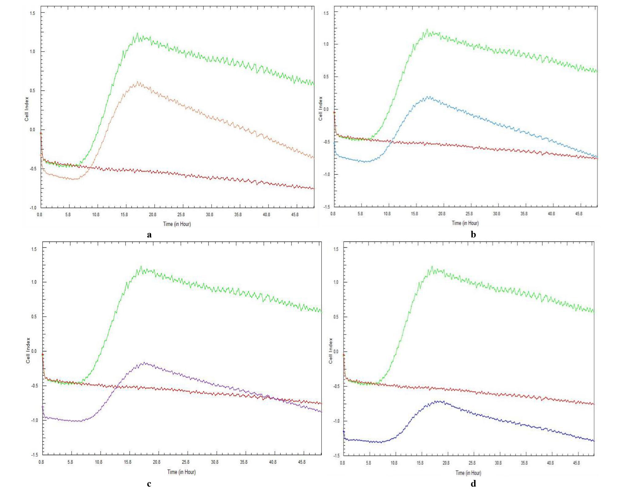
Figure 3. Real-time cell analysis cell index variations of Candida albicans (ATCC 90028) cell adhesion and subsequent biofilm formation in the presence of various concentrations of Galenia africana aqueous extract: a) 6.25 mg/mL, b) 12.5 mg/mL, c) 25 mg/mL, and d) 50 mg/mL. A positive control (green curve) and sterility control (burgundy curve) are included.
When testing C. glabrata, the lowest concentration of G. africana (Figure 4a) did not appear to prevent adhesion and biofilm formation, as adhesion was still evident with CI values similar to the positive control. Unlike C. albicans, in which an effect on the adhesion and CI value was observed at a G. africana concentration of 6.25 mg/mL, an effect on C. glabrata adhesion was only observed at a concentration of 12.5 mg/mL, where a significant difference to the control was observed. Concentrations of G. africana within the range of 12.5-50 mg/mL (Figure 4b-d) proved to be the most effective in reducing the CI values, while the 25 and 50 mg/mL concentrations of G. africana (Figure 4c and d) maintained negative C. glabrata CI values. The changes in the maximum CI values obtained for both the 10-h preformed biofilm disruption and biofilm prevention demonstrated a dose-dependent response of both Candida species to the G. africana extract, with the C. albicans maximum CI values being higher than those of C. glabrata (Table 1).
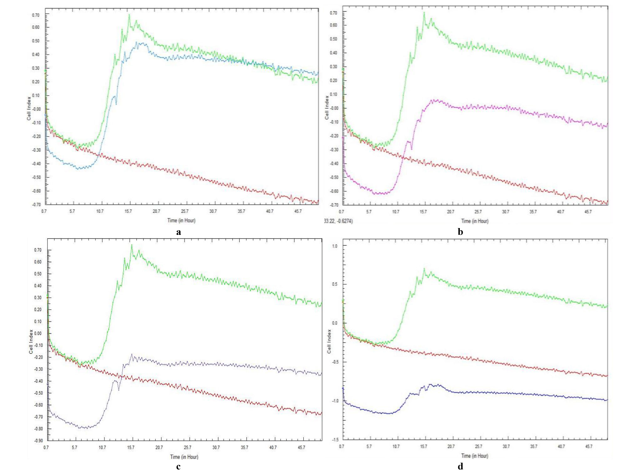
Figure 4. Real-time cell analysis cell index variations of Candida glabrata (ATCC 26512) cell adhesion and subsequent biofilm formation in the presence of various concentrations of Galenia africana aqueous extract: a) 6.25 mg/mL, b) 12.5 mg/mL, c) 25 mg/mL, and d) 50 mg/mL. A positive control (green curve) and sterility control (burgundy curve) are included.
| 10-hour preformed biofilm disruption maximum cell index values (after G. africana extract addition) | |||||
|---|---|---|---|---|---|
| Galenia africana concentration | 0 mg/mL | 6.25 mg/mL | 12.5 mg/mL | 25 mg/mL | 50 mg/mL |
| Candida albicans | 1.5 | 1.12 | 0.68 | 0.2 | -0.35 |
| Candida glabrata | 0.89 | 0.55 | 0.25 | -0.2 | -0.45 |
| Difference | 0.61 | 0.57 | 0.43 | 0.4 | 0.1 |
| Cell adhesion/biofilm prevention maximum CI values | |||||
| Galenia africana concentration | 0 mg/mL | 6.25 mg/mL | 12.5 mg/mL | 25 mg/mL | 50 mg/mL |
| Candida albicans | 1.24 | 0.62 | 0.2 | -0.15 | -0.72 |
| Candida glabrata | 0.69 | 0.49 | 0.06 | -0.18 | -0.78 |
| Difference | 0.55 | 0.13 | 0.14 | 0.03 | 0.06 |
Since this was a preliminary study showing the potential antibiofilm activity of G. africana extract against two Candida species, no statistical analysis was performed.
Discussion
This study aimed to monitor and assess the anti-biofilm properties of a G. africana ethanolic extract against the biofilm formation of C. albicans (ATCC 90028) and C. glabrata (ATCC 26512) in real-time, while simultaneously assessing the ability of the xCELLigence RTCA DP system to be used as a tool for screening novel medicinal plants. To the best of our knowledge, this is the first reported study to use an impedance-based system to monitor the real-time effects of a novel medicinal plant on Candida biofilm formation.
Although the xCELLigence system has been validated as an investigative tool for multiple complex cellular behaviors, medication responses [ 53 - 55 ], and monitoring of microbial biofilm formation [ 46 , 56 ], its use in real-time biofilm disruption using herbal extracts has not been previously documented.
There has been an increasing interest in the real-time monitoring of biofilm formation, moving away from traditional, end-point methods, such as crystal violet staining, the 2,3-bis(2-methoxy-4-nitro-5-sulfophenyl)-5-[phenylamino)carbonyl]-2H-tetrazolium hydroxide reduction assay, dry cell weight and viable cell counts, as well as molecular methods, such as DNA and protein quantification [ 57 - 60 ]. End-point methods allow for the observation or measurement of the final effect but they are labor intensive and require invasive sampling, with long interludes between sampling and obtaining a result. Conversely, the RTCA system monitors adhesion and biofilm formation in real time [ 56 ], giving a more in-depth understanding of each phase of biofilm formation [ 46 ]. In principle, the CI values increase as the biofilm adheres and proliferates on the bottom of the E-plate wells, with a peak CI being consistent with the start of biofilm maturation and a decrease occurring at the start of the biofilm detachment phase [ 56 ]. A declining slope after reaching the peak CI value has been suggested as a measure of biofilm formation [ 56 , 61 ], with further declining numbers associated with the death phase [ 62 ].
By allowing the biofilm of each Candida species to become established and subsequently treating them with varying concentrations of G. africana, biofilm disruption was achieved, although at relatively high concentrations (50 mg/mL for C. albicans and 25 mg/mL for C. glabrata). The response to G. africana was immediate and transient, as revealed by the precise timing and magnitude of the response by the xCELLigence system. This response could be due to the higher volume of flavanols/pinocembrin in higher doses, as they are the most active molecules within the G. africana extract [ 63 , 64 ].
In conventional in vitro assays, treatments are usually executed at predetermined time points that are convenient (e.g., 12 or 24 h post-seeding, depending on the cell type), rather than on experimental or behavioral data from the cells [ 45 ]. By contrast, the xCELLigence CI curve data revealed real-time information related to the behavior, growth, and overall health of the cells, which can be used as a guide to improve the experimental design [ 45 ].
Biofilm formation can be prevented by inhibition of its establishment [ 65 ]. In the present study, the lowest concentration of G. africana did not appear to prevent the adhesion and biofilm formation of C. albicans, as adhesion was still evident after the administration of 12.5 mg/mL extract with a decreased CI that remained negative after 24-hour incubation, while the two higher concentrations successfully inhibited C. albicans biofilm formation for the duration of the 48-hour incubation. In the case of C. glabrata, there was a short period of adhesion at a 12.5 mg/mL extract exposure, albeit at much lower CI values than C. albicans. This difference could be attributed to C. albicans being more strongly resistant in a sessile community, compared to a planktonic state [ 66 ]. Specific adhesins allow firm attachment of the microorganisms [ 67 ] and facilitate the formation of biofilms; therefore, according to the biofilm eradication and prevention curves, C. albicans had more adhesion potential than C. glabrata. The two higher concentrations completely prevented adhesion and subsequent biofilm formation for the duration of the 48-hour incubation period.
Khun et al. [ 68 ] found that C. albicans produced more biofilm, compared to NAC species, while Marak and Dhanashree [ 69 ] reported greater biofilm formation with C. parapsilosis (100%), C. tropicalis (61.53%), and C. krusei (55.55%), compared to C. albicans (39.02%), with C. glabrata showing no biofilm production. These studies support the adhesion capability and subsequent biofilm formation of C. albicans and C. glabrata in the present study, even though the methods and strains were different.
A previous study of biofilm formation of three C. albicans strains (one producing widespread biofilms and two mutant strains lacking adhesins) using several end-point techniques suggested that in vitro experiments may not accurately reflect in vivo realities [ 60 ]. Therefore, a comparison of strong biofilm-producing Candida cells and mutant strains defective in producing biofilms is desirable and overcomes a limitation of the current study.
Conclusion
The authors are confident that the study objectives were achieved since the xCelligence RTCA could demonstrate the ability of G. africana to prevent the formation of Candida biofilms and disrupt existing Candida biofilms.
Further studies using the the xCelligence RTCA could explore the antimicrobial effects that individual parts of the G. africana plant exhibit in susceptibly assays. It would also be beneficial to test other medication-resistant Candida species with various adherence properties, such as C. krusei. Studies investigating the protein expression of medicinal plants and correlating it with their mode of action are warranted before the elucidation of gene expression data.
Acknowledgments
The authors thankfully acknowledge Anatech Analytical Technology (Johannesburg, South Africa) for the use of the xCELLigence RTCA instrument and Christelle Klopper from Anatech Instruments for her excellent technical advice.
Authors’ contributions
J. K. and C. A. conceptualized the study and acquired funding, C. A. and J. K. provided resources, K. S. and P. A. developed the methodology, K. S. conducted the investigation, K. S. and P. A. handled visualization, K. S. prepared the original draft, and P. A., J. K., and C. A. contributed to review and editing. All authors gave their final approval and agreed to be accountable for all aspects of the work.
Conflicts of interest
The authors declare that they have no conflict of interest.
Financial Disclosure
This study was partially funded by the National Research Foundation of South Africa. Any opinions, findings, conclusions, and recommendations expressed in this manuscript are those of the authors, and therefore, the National Research Foundation does not accept any liability in regard thereto.
References
- Lortholary O, Renaudat C, Sitbon K, Madec Y, Denoeud-Ndam L, Wolff M, et al. Worrisome trends in incidence and mortality of candidemia in intensive care units (Paris area, 2002-2010). Intensive Care Med. 2014; 40(9):1303-12.
- Sasso M, Roger C, Sasso M, Poujol H, Barbar S, Lefrant JY, et al. Changes in the distribution of colonising and infecting Candida spp. isolates, antifungal drug consumption and susceptibility in a French intensive care unit: a 10-year study. Mycoses. 2017; 60(12):770-80.
- Montagna MT, Caggiano G, Lovero G, De Giglio O, Coretti C, Cuna T, et al. Epidemiology of invasive fungal infections in the intensive care unit: results of a multicentre Italian survey (AURORA Project). Infection. 2013; 41(3):645-53.
- Guo F, Yang Y, Kang Y, Zang B, Cui W, Qin B, et al. Invasive candidiasis in intensive care units in China: a multicentre prospective observational study. J Antimicrob Chemother. 2013; 68(7):1660-8.
- Puig-Asensio M, Peman J, Zaragoza R, Garnacho-Montero J, Martin-Mazuelos E, Cuenca-Estrella M, et al. Impact of therapeutic strategies on the prognosis of candidemia in the ICU. Crit Care Med. 2014; 42(6):1423-32.
- Ahmad A, Husain A, Khan S, Mujeeb M, Bhandari A. Design, synthesis, molecular properties and antimicrobial activities of some novel 2(3H) pyrrolone derivatives. J Saudi Chem Soc. 2015; 19(3):340-6.
- Hoenigl M, Salmanton-García J, Egger M, Gangneux JP, Bicanic T, Arikan-Akdagli S, et al. Guideline adherence and survival of patients with candidaemia in Europe: results from the ECMM Candida III multinational European observational cohort study. Lancet Infect Dis. 2023; 23(6):751-61.
- Katsipoulaki M, Stappers MHT, Malavia-Jones D, Brunke S, Hube B, Gow NAR. Candida albicans and Candida glabrata: global priority pathogens. Microbiol Mol Biol Rev. 2024; 88(2):e0002123.
- Timmermans B, De Las Peñas A, Castaño I, Van Dijck P. Adhesins in Candida glabrata. J Fungi (Basel). 2018; 4(2):60.
- Brown ED, Wright GD. Antibacterial drug discovery in the resistance era. Nature. 2016; 529(7586):336-43.
- Fairlamb AH, Gow NA, Matthews KR, Waters AP. Drug resistance in eukaryotic microorganisms. Nat Microbiol. 2016; 1(7):1-33.
- Robbins N, Wright GD, Cowen LE. Antifungal Drugs: The current armamentarium and development of new agents. Microbiol Spectr. 2016; 4(5):1-20.
- Cornely OA, Bassetti M, Calandra T, Garbino J, Kullberg BJ, Lortholary O, et al. ESCMID* guideline for the diagnosis and management of Candida diseases 2012: non-neutropenic adult patients. Clin Microbiol Infect. 2012; 18(Suppl 7):19-37.
- Elhoufi A, Ahmadi A, Asnaashari AM, Davarpanah MA, Bidgoli BF, Moghaddam OM, et al. Invasive candidiasis in critical care setting, updated recommendations from “Invasive Fungal Infections-Clinical Forum”, Iran. World J Crit Care Med. 2014; 3(4):102-12.
- Pappas PG, Kauffman CA, Andes DR, Clancy CJ, Marr KA, Ostrosky-Zeichner L, et al. Clinical practice guideline for the management of candidiasis: 2016 update by the Infectious Diseases Society of America. Clin Infect Dis. 2016; 62(4):e1-50.
- Bienvenu AL, Pradat P, Guerin C, Aubrun F, Fellahi JL, Friggeri A, et al. Evaluation of first-line therapies for the treatment of candidemia in ICU patients: a propensity score analysis. Int J Infect Dis. 2020; 93:15-21.
- Perlin DS. Echinocandin Resistance in Candida. Clin Infect Dis. 2015; 61(Suppl 6):S612-7.
- Pham CD, Iqbal N, Bolden CB, Kuykendall RJ, Harrison LH, Farley MM, et al. Role of FKS mutations in Candida glabrata: MIC values, echinocandin resistance, and multidrug resistance. Antimicrob Agents Chemother. 2014; 58(8):4690-6.
- Perlin DS. Mechanisms of echinocandin antifungal drug resistance. Ann N Y Acad Sci. 2015; 1354(1):1-11.
- Hull CM, Bader O, Parker JE, Weig M, Gross U, Warrilow AGS, et al. Two clinical isolates of Candida glabrata exhibiting reduced sensitivity to amphotericin B both harbor mutations in ERG2. Antimicrob Agents Chemother. 2012; 56(12):6417-21.
- Thompson III GR, Wiederhold NP, Vallor AC, Villareal NC, Lewis JS, Patterson TF. Development of caspofungin resistance following prolonged therapy for invasive candidiasis secondary to Candida glabrata infection. Antimicrob Agents Chemother. 2008; 52(10):3783-5.
- Fekkar A, Dannaoui E, Meyer I, Imbert S, Brossas JY, Uzunov M, et al. Emergence of echinocandin-resistant Candida spp. in a hospital setting: a consequence of 10 years of increasing use of antifungal therapy? Eur J Clin Microbiol Infect Dis. 2014; 33(9):1489-96.
- Ramage G, Rajendran R, Sherry L, Williams C. Fungal biofilm resistance. Int J Microbiol. 2012; 2012:1-14.
- Rautemaa R, Ramage G. Oral candidosis – clinical challenges of a biofilm disease. Crit Rev Microbiol. 2011; 37(4):328-36.
- Pierce CG, Uppuluri P, Tristan AR, Wormley FL, Mowat E, Ramage G, et al. A simple and reproducible 96-well plate-based method for the formation of fungal biofilms and its application to antifungal susceptibility testing. Nat Protoc. 2008; 3(9):1494-500.
- Donlan RM. Biofilm formation: a clinically relevant microbiological process. Clin Infect Dis. 2001; 33(8):1387-92.
- Mukherjee PK, Chandra J, Kuhn DM, Ghannoum MA. Mechanisms of azole resistance in Candida albicans biofilms: phase-specific role of efflux pumps and membrane sterols. Infect Immun. 2003; 71(8):4333-40.
- Shahzad M, Sherry L, Rajendran R, Edwards CA, Combet E, Ramage, G. Utilizing polyphenols for the clinical management of Candida albicans biofilms. Int J Antimicrob Agents. 2014; 44(3):269-73.
- Gulati M, Nobile CJ. Candida albicans biofilms: development, regulation, and molecular mechanisms. Microbes Infect. 2016; 18(5):310-21.
- Nett JE, Andes DR. Contributions of the biofilm matrix to Candida pathogenesis. J Fungi (Basel). 2020; 6(1):21.
- Tumbarello M, Posteraro B, Trecarichi EM, Fiori B, Rossi M, Porta R, et al. Biofilm production by Candida species and inadequate antifungal therapy as predictors of mortality for patients with candidemia. J Clin Microbiol. 2007; 45(6):1843-50.
- Wall G, Montelongo-Jauregui D, Vidal Bonifacio B, Lopez-Ribot JL, Uppuluri P. Candida albicans biofilm growth and dispersal: contributions to pathogenesis. Curr Opin Microbiol. 2019; 52:1-6.
- World Health Organization. WHO fungal priority pathogens list to guide research, development and public health action. Geneva: WHO; 2022.
- Kabir MA, Hussain MA, Ahmad Z. Candida albicans: a model organism for studying fungal pathogens. ISRN Microbiol. 2012; 2012:1-15.
- Afolayan AJ, Lewu FB. Antimicrobial activity of Alepidea amatymbica. Pharm Biol. 2009; 47:436-9.
- Samber N, Khan A, Varma A, Manzoor N. Synergistic anti-candidal activity and mode of action of Mentha piperita essential oil and its major components. Pharm Biol. 2015; 53(10):1496-504.
- Armengol ES, Harmanci M, Laffleur F. Current strategies to determine antifungal and antimicrobial activity of natural compounds. Microbiol Res. 2021; 252:1-12.
- Twilley D, Rademan S, Lall N. A review on traditionally used South African medicinal plants, their secondary metabolites and their potential development into anticancer agents. J Ethnopharmacol. 2020; 261:1-17.
- Mudau FN, Chimonyo VGP, Modi AT, Mabhaudhi T. Neglected and underutilised crops: a systematic review of their potential as food and herbal medicinal crops in South Africa. Front Pharmacol. 2022; 12(809866):1-31.
- Kellerman TS, Coetzer JAW, Naude TW. Plant poisoning and mycotoxicoses of livestock in Southern Africa. Cape Town, South Africa: Oxford University Press; 1988.
- Vries FA, El Bitar H, Green IR, Klaasen JA, Mabulesa WT, Bodo B, et al. An antifungal active extract from the aerial parts of Galenia africana. 11th NAPRECA symposium book of proceedings, Antananarivo, Madagascar. 2005;123-31.
- Ng’uni TL, dos Santos Abrantes PM, McArthur C, Klaasen JA, Fielding BC. Evaluation of synergistic anticandidal activity of Galenia africana extract and fluconazole against Candida albicans and Candida glabrata. J Herb Med. 2022; 32:100503.
- Dias LG, Meirinho SG, Veloso AC, Rodrigues LR, Peres AM. Electronic tongues and aptasensors. InBioinspired Materials for Medical Applications. 2017;371-402.
- Junka AF, Janczura A, Smutnicka D, Mączyńska B, Anna S, Nowicka J, et al. Use of the real time xCELLigence system for purposes of medical microbiology. Pol J Microbiol. 2012; 61(3):191-7.
- Kho D, MacDonald C, Johnson R, Unsworth CP, O'Carroll SJ, du Mez E, et al. Application of xCELLigence RTCA biosensor technology for revealing the profile and window of drug responsiveness in real time. Biosensors. 2015; 5(2):199-222.
- Ferrer MD, Rodriguez JC, Alvarez L, Artacho L, Royo G, Mira A. Effect of antibiotics on biofilm inhibition and induction measured by real-time cell analysis. J Appl Microbiol. 2017; 122(3):640-50.
- Hamidi H, Lilja J, Ivaska J. Using xCELLigence RTCA instrument to measure cell adhesion. Bio-protoc. 2017; 7(24):1-16.
- Alshanta OA, Shaban S, Nile CJ, McLean W, Ramage G. Candida albicans biofilm heterogeneity and tolerance of clinical isolates: Implications for secondary endodontic infections. Antibiotics (Basel). 2019; 8(4):204.
- Abrantes PM, Africa CW. Measuring Streptococcus mutans, Streptococcus sanguinis and Candida albicans biofilm formation using a real-time impedance-based system. J Microbiol Methods. 2020; 169:1-5.
- Abrantes PM, Behardien K, Africa CW. Real-time assessment of interspecies Candida biofilm formation. Open Microbiol J. 2022; 16:1-8.
- Leonhard M, Zatorska B, Moser D, Tan Y, Schneider-Stickler B. Evaluation of combined growth media for in vitro cultivation of oropharyngeal biofilms on prosthetic silicone. J Mater Sci Mater Med. 2018; 29(4): 45.
- Dunn MJ, Fillinger RJ, Anderson LM, Anderson MZ. Automated quantification of Candida albicans biofilm-related phenotypes reveals additive contributions to biofilm production. NPJ Biofilms Microbiomes. 2020; 6(1):36.
- Martinez-Serra J, Gutierrez A, Muñoz-Capó S, Navarro-Palou M, Ros T, Amat JC, et al. xCELLigence system for real-time label-free monitoring of growth and viability of cell lines from hematological malignancies. Onco Targets Ther. 2014; 7:985-94.
- Urcan E, Haertel U, Styllou M, Hickel R, Scherthan H, Reichl FX. Real-time xCELLigence impedance analysis of the cytotoxicity of dental composite components on human gingival fibroblasts. Dent Mater J. 2010; 26(1):51-8.
- Guan N, Deng J, Li T, Xu X, Irelan JT, Wang MW. Label-free monitoring of T cell activation by the impedance-based xCELLigence system. Mol Biosyst. 2013; 9(5):1035-43.
- Gutiérrez D, Hidalgo-Cantabrana C, Rodríguez A, García P, Ruas-Madiedo P. Monitoring in real time the formation and removal of biofilms from clinical related pathogens using an impedance-based technology. PloS One. 2016; 11(10):1-17.
- Chandra J, Mukherjee PK, Leidich SD, Faddoul FF, Hoyer LL, Douglas LJ, et al. Antifungal resistance of candidal biofilms formed on denture acrylic in vitro. J Dent Res. 2001; 80(3):903-8.
- Chandra J, Kuhn DM, Mukherjee PK, Hoyer LL, McCormick T, Ghannoum M. Biofilm formation by the fungal pathogen Candida albicans: development, architecture, and drug resistance. J Bacteriol. 2001; 183(18):5385-94.
- Jin Y, Yip HK, Samaranayake YH, Yau JY, Samaranayake LP. Biofilm-forming ability of Candida albicans is unlikely to contribute to high levels of oral yeasts carriage in cases of Human Immunodeficiency Virus infection. J Clin Microbiol. 2003; 41(7):2961-7.
- Taff HT, Nett JE, Andes DR. Comparative analysis of Candida biofilm quantitation assays. Med Mycol. 2012; 50(2):214-8.
- van Duuren JBJH, Müsken M, Karge B, Tomasch J, Wittmann C, Häussler S, et al. Use of single-frequency impedance spectroscopy to characterize the growth dynamics of biofilm formation in Pseudomonas aeruginosa. Sci Rep. 2017; 7(1):1-12.
- Garrett TR, Bhakoo M, Zhang Z. Bacterial adhesion and biofilms on surfaces. Prog Nat Sci. 2008; 18(9):1049-56.
- Mativandlela SPN, Muthivhi T, Kikuchi H, Oshima Y, Hamilton C, Hussein AA, et al. Antimycobacterial flavonoids from the leaf extract of Galenia africana. J Nat Prod. 2009; 72(12):2169-71.
- Ticha LA, Klaasen JA, Green IR, Naidoo S, Baker B, Pietersen RD. Phytochemical and antimicrobial screening of flavanones and chalcones from Galenia africana and Dicerothamnus rhinocerotis. Nat Prod Commun. 2015; 10(7):1185-90.
- Friedlander A, Nir S, Reches M, Shemesh M. Preventing biofilm formation by dairy-associated bacteria using peptide-coated surfaces. Front Microbiol. 2019; 10:1405.
- Ramage G, López-Ribot JL. Techniques for antifungal susceptibility testing of Candida albicans biofilms. Methods Mol Med. 2005; 118:71-9.
- Pugazhendhi AS, Wei F, Hughes M, Coathup M. Bacterial adhesion, virulence, and biofilm formation. InMusculoskeletal Infection. 2022;19-64.
- Khun DM, Chandra J, Mukherjee PK, Ghannoum, MA. Comparison of biofilms formed by Candida albicans and Candida parapsilosis on bioprosthetic surfaces. Infect Immun. 2002; 70(2):878-88.
- Marak MB, Dhanashree B. Antifungal susceptibility and biofilm production of Candida spp. isolated from clinical samples. Int J Microbiol. 2018; 2018:1-5.


