
Introduction
Fungal respiratory infections represent an escalating concern and a major cause of mortality in immunocompromised patients [ 1 , 2 ]. The most common fungal pneumonia etiological agents include Aspergillus, Mucorales, Cryptococcus, and endemic dimorphic fungi. [ 3 , 4 ]. The genus Aspergillus from the division Ascomycota is among the most prevalent and opportunistic molds worldwide [ 5 ]. The attributable mortality rate of pneumonia caused by Aspergillus is more than 80% [ 6 ]. Several species of Aspergillus spp., notably A. fumigatus, A. flavus, and A. niger, have been documented to cause respiratory diseases in humans [ 7 - 9 ]. Aspergillus spp. can release millions of spores daily, which can induce infections and chronic pulmonary disease in humans via inhalation [ 10 ]. These spores produced by fungi can survive for extended durations until environmental conditions become conducive for growth. It should be mentioned that dispersing these spores may lead to aspergillosis in susceptible individuals [ 11 ].
As a commonly utilized strategy, treatment using antifungal agents (such as itraconazole and ketoconazole) has some limitations due to systemic antibiotic consumption, the instability of oral drugs in aqueous intestinal fluids, and the main first-pass metabolism of CYP3A before entering the systemic circulation [ 12 , 13 ]. All of these drawbacks are responsible for significant adverse effects and low bioavailability of the agents. Despite extensive investigations into the pathogenesis and prevention of pulmonary aspergillosis, as well as efforts to apply prophylactic interventions targeting this infection (e.g., regulation of the tracheal tube cuff and reduced sedation protocols), the burden of fungal infections remains a major concern in patients undergoing ventilation within intensive care units [ 14 ]. Given that the clinical presentation of pneumonia arises over several days following the establishment of infection, with incidence peaking around 7 days after the initiation of mechanical ventilation, a therapeutic window exists to curtail infectious colonization and precocious spread in the intervening period [ 15 ]. To overcome the problems associated with traditional treatment, pulmonary delivery of nano-formulations of antifungal drugs have become a focal point of research due to the advantages of targeted delivery.
Nanoparticles, with their tunable size and surface properties, can be engineered to enhance drug delivery directly to the site of infection within the alveolar space. This targeted approach facilitates increased drug concentrations at the fungal infection site, reduces systemic exposure, improves bioavailability, leads to slower clearance, and prolongs therapeutic effects. Furthermore, surface modification of nanoparticles can enable specific interactions with alveolar cells or the fungal pathogens themselves, enhancing drug uptake and antifungal activity. Consequently, nanoparticulate drug carriers represent a promising strategy for overcoming the challenges associated with treating pulmonary infections and improving patient outcomes.
Nanocarriers for pulmonary delivery against fungal infection
Nanoparticles can serve as effective pharmaceutical delivery platforms for treating pulmonary mycoses due to their great surface area-to-volume proportion and capacity to augment the bioactivity of anti-mycotic drugs [ 16 - 18 ]. Given their typically small dimensions (below 100 nanometers), these formulations can permeate into alveolar cavities, thereby facilitating optimal anti-mycotic localization at the sites of pulmonary infection [ 19 , 20 ]. Additionally, these nano-systems exhibit augmented permeation and retention, which can be useful for targeted delivery and controlled release via increased solubility, protection against the degradation of enzymes, enhanced blood circulation time, and increased absorption by epithelial cells [ 21 , 22 ]. These properties enable lower doses of antifungal drugs to be administered, thereby reducing whole-body exposure to the pharmacological agents, which can potentially decrease the risk of adverse reactions linked to azole therapy, such as hepatotoxicity [ 23 , 24 ]. Nanocarriers may consist of lipid or polymeric constituents, but lipid-based platforms are preferentially considered due to their greater physiological compatibility and biodegradability over time [ 25 ]. Nevertheless, a drug transportation system capable of surmounting pulmonary barriers is imperative to optimize the efficacy of pharmaceutical agents within respiratory tract epithelial cells.
In a promising study, lipid-polymer hybrid nanoparticles were coupled with nucleic acid cargo to tackle cystic fibrosis mucus secretions [ 26 ]. Results of the aforementioned study confirmed the ability of non-PEGylated lipid-polymer hybrid nanoparticles to overcome mucus barriers in the airway epithelium. Notably, PEGylation did not enhance mucus permeation in sticky and complex samples. Liposomal amphotericin B (AmBisome) was named the first U. S. Food and Drug Administration-approved polyene antifungal drug [ 27 ]. As a result of nanoliposome encapsulation, the pharmacokinetics of amphotericin B were altered, with prolonged circulation and selective transfer into fungal cells, thereby reducing nephrotoxicity. However, the conventional amphotericin B liposomal nano-formulation has a tendency for antifungal agent leakage from the carrier during blood circulation, major clearance of it by the reticuloendothelial system, and incapacity for extravasation into the infected epithelial cells.
In a recent study, it has been suggested that three preparation methods, including a solvent-free approach, a solvent evaporation procedure, and ethanol injection, be used to encapsulate amphotericin B in liposomes [ 28 ]. Valuable advantages of the solvent-free method include the removal of the evaporation stage, a major concern for industrial-scale manufacturers, and the production of a harmless final product in biosystems. The assessment based on transmission electron microscopy analysis indicated that after the lyophilization process, the liposomal amphotericin B preserved its spherical nanostructure without aggregation or deformations. In addition, storage in the refrigerator can enhance the stability of this powder. In contrast, previous articles have shown that liposomal amphotericin B is unstable over long periods in solution form, as it undergoes photolytic degradation, oxidation, and hydrolysis [ 29 ]. However, in the solvent-free procedure, after lyophilization, the size of the nanoparticles increased but remained within the size range considered appropriate for nano-liposomal delivery systems [ 29 ].
Table 1 presents a comparative analysis of four categories of nanomaterials for fungal infections.
| Characteristics | Nano-graphene Oxide [ 31 - 33 ] | Liposomes [ 34 - 36 ] | Hybrid Nanoparticles [ 37 - 39 ] | Nano Metallic particles [ 40 - 42 ] |
|---|---|---|---|---|
| Composition | Graphene with oxygen-containing groups | Phospholipids and cholesterol | Lipid and polymer | Metals and metal oxides |
| Preparation | Oxidation of graphene | Encapsulation of drugs in spherical bilayers | Combination of lipid and polymer | Chemical or biological synthesis of metals |
| Production Cost | Moderate | Moderate | Variable | High |
| Shape characteristics | Nanoscale-thin sheets | Spherical lipid bilayers | Diverse shapes with functional groups | Various shapes depending on the type of metal |
| Particle Size | 100-200 nm | 50-300 nm | 50-200 nm | 1-100 nm |
| Stability | Unstable, requires lyophilization | Drug leakage possible | Good stability | Stability depends on the metal type |
| Mechanism of action in fungal infection treatment | ROS, pore formation in fungal walls (nano-knives), fungal cell encirclement blocking access to oxygen and nutrients, and biofilm inhibition. Synergistic effects when combined with certain antifungal drugs | Reduction of toxicity and enhancement of antifungal drug penetration | Enhancement of drug absorption and penetration to deep tissues | Oxidative stress, membrane disruption |
| Drug delivery capability | Drug delivery to the infection site, reducing side effects, and inducing targeted delivery | Improvement of drug delivery and release | Enhancement of drug transport to the injection site | Enhancement of drug delivery to the infection site |
| Characteristics | NGO | Liposomes | Hybrid Nanoparticles | Other Nanoparticles (Metallic) |
| Safety and biocompatibility | Toxicity at high concentrations, surface modification improves biocompatibility | Liposomal drug forms are less toxic than traditional forms | Usage of biocompatible materials can reduce toxicity | The variable depends on the metal and its preparation |
| Administration method | Inhalation | Mostly intravenous | Inhalation possible | Variable |
| Stability in solution | It may be unstable in solution, requires lyophilization | It may be unstable in solution, prone to leakage | Better stability, compared to simple nanoparticles | Variable |
| Penetration capability | Small size enables deep penetration into the lungs and infection sites | Limited penetration into deep tissues | Special coatings improve deep tissue penetration | Variable |
| Impact on the immune system | It may induce inflammation in some cases | Issues, like drug leakage and low stability | Compared to simple nanoparticles, better cell uptake, lung retention, and antifungal effects | Variable |
| Surface area | High | Moderate | Variable | Variable |
| Surface modifiability | High | Moderate | High | Moderate |
| NGO: nano-graphene oxide, ROS: reactive oxygen species | ||||
Challenges associated with available anti-fungal liposomes
Up to now, several nano-liposomal antifungal formulations (such as amphotericin B) have been developed; however, these nano-formulations still suffer from problems, such as stability, lipid peroxidation, and cellular toxicity caused by nontargeted delivery of liposomes via the parenteral route [ 30 ]. In addition, the rapid removal of these nanoparticles from blood circulation caused by uptake by the reticuloendothelial system, with significant decomposition in the liver, is another factor that cannot be overlooked.
Notably, the treatment of fungal infections that are situated in the lower respiratory tract or penetrate further into the lung parenchyma can prove challenging [ 43 ]. This is due to the fact that maintaining therapeutically effective concentrations of an antifungal agent at the site of infection over an extended duration is often impossible. Rapid clearance of drugs from these highly vascularized pulmonary regions, where blood flow turnover is elevated, can limit the sustained presence of sufficient drug levels needed for optimal treatment efficacy. Locating infections in deeper recesses of the lung that are perfused by profuse circulation renders fungal therapeutic interventions more difficult.
In this regard, a nanoliposome-based inhaled system was designed to directly deliver antifungal drugs to the lungs, which can reduce drug levels throughout the blood while maintaining therapeutic concentrations at the focal of infection, thereby reducing nephrotoxicity, compared to intravenous administration. Inhalation therapy is employed for the local treatment of pulmonary diseases, including chronic obstructive pulmonary disease, pneumonia, bronchiectasis, asthma, cystic fibrosis, and aspergillosis.
Local inhalation of drugs targets the lungs for therapeutic effects, offering several advantages over other administration routes, including a large absorption area, rapid absorption due to a thin membrane, reduced enzyme activity that bypasses the first-pass effect of the liver, and the ability to maintain high drug concentrations at the target site. This method presents a non-invasive alternative to injections, allowing for lower doses and enhanced bioavailability [ 44 ].
Extensive research has indicated that common antimicrobial drugs, such as amphotericin B, amikacin, tobramycin, and rifampicin, which are often used to combat lower respiratory infections caused by pathogens, such as Mycobacterium tuberculosis, Candida albicans, and Pseudomonas aeruginosa, have serious side effects when administered intravenously or orally. However, when administered via inhaled drug delivery, their side effects are significantly ameliorated [ 45 , 46 ].
A broad array of pulmonary fungal diseases, encompassing invasive aspergillosis, chronic pulmonary aspergillosis, Aspergillus bronchitis, and allergic bronchopulmonary aspergillosis, is predominantly attributed to Aspergillus spp., especially A. fumigatus. Regardless of their clinical manifestations, azoles serve as the first-line therapeutic approach [ 47 ].
Pulmonary inhalation is useful for drugs with limited oral absorption, such as amphotericin B, due to its low water solubility and low membrane permeability [ 45 ]. Additionally, due to the low metabolic activity in the cellular lung, pulmonary inhalation delivery is important for the delivery of medications that are sensitive to gastric conditions, such as pH and enzymatic activity, especially those prone to hepatic metabolism [ 45 ]. This parallels research demonstrating that liposomal dry powder inhalation aerosols designed for targeted lung delivery are advantageous for compounds vulnerable to gastrointestinal and liver enzymatic breakdown.
In theory, inhaled liposome formulations could provide sufficiently high concentrations at the site of infection that exceed the minimum inhibitory concentration (MIC) for multidrug-resistant pathogens and, therefore, reduce the likelihood of developing drug resistance [ 45 ]. As a result, inhaled liposome antibiotics may limit opportunities for pathogens to evolve strategies of resistance, since low concentrations below the MIC of a drug could lead to increased mutation rates and, hence, the emergence of resistant subpopulations of pathogens that cannot be eradicated. However, meticulous attention must be paid to formulation engineering to improve consistent bioavailability, as design considerations can significantly impact this parameter.
Various factors, such as the dissolution of antifungal aerosol nanoparticles in mucus and epithelial fluid, the appropriate diffusion of the formulation toward microorganisms at the mucosal site, the ability of the antibiotic against resistant mucoidal strains, and diffusion into spaces containing sputum, affect the clinical effectiveness of inhaled liposome antibiotics, especially in patients with cystic fibrosis. Delivery of liposomes via nebulization can lead to alterations in their physicochemical characteristics, and the deposition of the formulation within pulmonary alveolar macrophages may influence their functions [ 48 ]. In addition, the pulmonary branching structure impedes access to deeper lung regions, while clearance mechanisms efficiently remove inhaled drugs [ 45 ]. Given that most chronic lung infections occur in the airways of the lung lumen, effective delivery of pulmonary drugs requires advanced aerosol formulation methodologies and multifaceted delivery systems. Therefore, administering treatments directly to the lungs is a challenge due to the intricate anatomical and physiological architecture of the respiratory tract.
Comparison of nanoliposome-based inhaled systems and inhaled hybrid nanoparticles
Many studies compared the performance of hybrid nanoparticles and that of plain nanoparticles, which have been used in pulmonary antibiotic delivery [ 49 ]. Results of various investigations have demonstrated that hybrid nanoparticles have greater cellular uptake, improved lung retention, and enhanced antifungal efficacy, compared to plain nanoparticles as a platform for pulmonary drug delivery [ 50 , 51 ].
In a study, nebulized hybrid nanoparticles (dipalmitoyl phosphatidylcholine-coated chitosan nanoparticles) of voriconazole were evaluated for pulmonary aspergillosis, and the results were compared with those of the plain drug and nanoparticles [ 52 ]. Coating nanoparticles with the lung-specific phospholipid dipalmitoyl phosphatidylcholine exhibited augmented biocompatibility and suitable interaction with lung surfactant monolayers since this phospholipid constitutes approximately 50-60% of pulmonary surfactant [ 53 ].
Furthermore, the presence of this phospholipid minimizes macrophage cell uptake, thereby facilitating nano-formulation diffusion and internalization into the alveolar epithelial cells. In addition, modification of chitosan nanoparticles with lung surfactant lipid, dipalmitoyl phosphatidylcholine, can be postulated to hinder the adhesion of nano-formulation with the mucus barrier and escape deposition in the upper respiratory system [ 54 ]. This is followed by nanoparticle integration with pulmonary surfactant monolayers, which leads to interaction with alveolar epithelial cells and improves their internalization via cellular pathways, thereby enhancing the residence time of the drug in the lungs. Findings of the present study confirmed previous reports demonstrating heterodispersity arising from the generated aerosol cloud [ 55 , 56 ]. However, it was noteworthy that the nanoparticles exhibited good size and structural integrity preservation within the nebulized droplets, without any discernible aggregation. Although the researchers claimed that this hybrid nanoparticle accumulated in the cytoplasm of pulmonary epithelial cells, evaluation of cellular uptake analyses revealed that internalization of lipid-polymer hybrid nanoparticles in Calu-3 and A549 cell lines increased with dose and time.
Nano graphene-based materials
After the discovery of graphene, nano-graphene oxides (NGOs) received unique attention to achieve better attributes in comparison to their counterparts [ 57 ]. The functional groups (hydroxyl, carbonyl, and epoxy) on the surface of NGO provide the potential for utilization in different biological applications, including antifungal, antibacterial, bioimaging, tissue engineering, drug delivery, and antimicrobial applications [ 58 , 59 ]. The outstanding solubility of NGOs in different solvents can be attributed to the presence of these functional groups on the surface of GO nanosheets [ 60 ].
NGO can be functionalized with a diverse range of biomolecules, including antibodies, genes, DNA, and RNA, to enhance their targeted delivery to the intended site of action [ 61 ]. Notably, it was observed that they exhibited potent antimicrobial activity through the mechanism of oxidative stress [ 62 ]. Table 2 summarizes some studies related to the effects of nanographene on fungi. Graphene and its derivatives are antimicrobial agents that can be utilized in the treatment of antibiotic-resistant infectious diseases, and they are considered safe for use in humans. NGOs can show a variety of antimicrobial activities, including antifungal properties, as discussed in the subsequent sections. Table 3 shows the effect of nano-graphene oxide in the treatment of fungal infections.
| References | Type of nanoparticle | Type of fungi | Fungal infections or target organs | Main results |
|---|---|---|---|---|
| Wagner et al. 2017 [ 63 ] | Polyethylene glycol-functionalized poly (lactic acid-co-glycolic acid) and graphene oxide nanoparticles | Candida albicans | Vaginal Candidiasis | Nanoparticle drug-delivery vehicles may exacerbate inflammation in active yeast vaginal infections by increasing inflammatory recruitment. |
| Diez-Orejas et al. 2018 [ 64 ] | GO | C. albicans | Systemic Candidiasis (bloodstream) | GO uptake by macrophages modulates their phagocytic capability and increases the clearance of dead microorganisms during infection. |
| Nguyen et al. 2019 [ 7 ] | G and GO | Aspergillus niger and Aspergillus flavus | Aspergillosis (Lungs, Sinuses) | Examined the toxic effects of graphene and graphene oxide on A. niger and A. flavus, revealing a 62% reduction in biomass and altered hyphae, with A. niger being more affected by GO and A. flavus by G. |
| Kahsay et al. 2020 [ 65 ] | GO | Trichophyton mentagrophytes and C. albicans | Dermatophytosis (Skin, Nails) and Candidiasis (Oral, Vaginal) | rGO/Fe3O4 NCs showed antifungal activities against T. mentagrophytes and C. albicans |
| Wang et al. 2021 [ 66 ] | GO | Fusarium graminearum | Fusarium Head Blight (Cereal Crops, Agricultural) | The strong synergistic activity of GO with existing fungicides demonstrated the great application potential of GO in pest management. |
| Zhang et al. 2022 [ 67 ] | GO | Bipolaris sorokiniana | Leaf spot disease (plants) | Inhibitory effect of GO on B. sorokiniana was primarily related to the destruction of the cell membrane. |
| Gottardo et al. 2023 [ 68 ] | Reduced graphene oxide (rGO) | Candida species, Epidermophyton, Microsporum, and Trichophyton species (Dermatophyte agents) | Candidiasis (nails, skin) and Dermatophytosis (skin, nails, scalp) | Both yeast and dermatophytes clinical isolates were inhibited at a minimum of 6 and 24 h, respectively, but after 2 and 12 h, they had initial antifungal interference, respectively |
| Selvi et al. 2024 [ 69 ] | Silver-decorated graphene oxide (Ag/GO) | Candida albicans, Candida krusei, and Candida tropicalis | Systemic and cutaneous candidiasis | In-vitro antifungal activity of Ag/rGO nanocomposite revealed a higher zone of inhibition against C. albicans. |
| Graphene oxide (GO), Reduced graphene oxide (rGO) | ||||
| Advantages | Mechanism of Action | Application |
|---|---|---|
| Rapid action and reduced drug usage | Forms a protective layer on the skin and inhibits fungal growth. | Treatment of superficial fungal infections |
| Reduced side effects | Delivers drugs directly to infection sites and enhances drug efficacy. | Treatment of systemic fungal infections |
| Early detection of infections | Acts as a contrast agent in imaging techniques. | Diagnosis of fungal infections |
| G: graphene, GO: graphene oxide | ||
Factors affecting the antifungal activity of nano-graphene oxide
In most NGO studies, notable distinctions have been observed in the antifungal activities between NGO nanosheets and graphite [ 70 , 71 ]. The significant antifungal effects of this material depend heavily on the concentration and duration of exposure [ 72 ]. Furthermore, as the percentage of functional groups increases and the size decreases, the toxicity effect increases, leading to a higher impact on fungal killing [ 73 , 74 ]. Research findings have indicated that while conductive reduced graphene oxide exhibits a significantly higher oxidation capacity, it is NGO layers with smaller lateral size dimensions that demonstrate superior antifungal properties. Moreover, the thickness of NGO layers and the angle of orientation with microorganisms may affect the anti-microbial efficiency [ 75 ]. Table 4 tabulates the characteristics of the NGO used in the treatment of fungal infections.
| Disadvantages | Advantages |
|---|---|
| Potential toxicity: At high concentrations, NGO can be toxic to human cells [ 76 ] | Strong antifungal effects: oxidative stress, membrane damage, fungal encapsulation, inhibition of fungal proliferation, and biofilm disruption [ 62 ] |
| Structural changes: An NGO may exhibit structural changes when exposed to environmental conditions, such as photodegradation, oxidation, and hydrolysis, which can affect its performance [ 77 ] | Drug delivery capability: enhances drug efficacy, reduces side effects, and allows for lower drug doses [ 78 ] |
| Accumulation and inflammation: Administration methods, like intratracheal injection or inhalation, may lead to NGO accumulation and increased inflammation in the lungs [ 77 ] | Therapeutic targeting enables specific penetration to the infection site. Inhalation can directly deliver the drug to the lungs and minimize systemic side effects [ 79 ] |
| Instability: The NGO may be unstable in solution and require lyophilization to maintain its structure [ 77 ] | Overcoming drug resistance: It increases drug concentration at the infection site, reducing opportunities for pathogen mutation and aiding in overcoming drug resistance [ 80 ]. |
| Body clearance: The NGO may be rapidly cleared from the bloodstream by the reticuloendothelial system and accumulate in the liver [ 81 ] | Enhanced biocompatibility: Surface modifications (e.g., polymer coatings) can increase the biocompatibility of the NGO [ 82 ] |
| Uncertainty in mechanisms: The Exact mechanisms of NGO action in the respiratory system are not fully understood [ 79 ] | High modifiability: NGOs, with various functional groups, can be modified and conjugated with biomolecules, such as antibodies, genes, and antifungal drugs [ 83 ] |
| Industrial production challenges: Industrial-scale production of NGO with consistent and desirable properties still faces challenges [ 84 ] | Synergistic effect: The Combination of NGO with antifungal drugs can create synergistic effects, enhancing therapeutic efficacy. Reduced drug dosage: It allows for lower drug doses, reducing side effects [ 83 ] |
Antifungal mechanisms of nano-graphene oxide
It has been reported that the NGO demonstrates favorable inhibitory effects against fungi (Figure 1). This activity is likely attributed to the ability of NGOs to disrupt microorganism membranes through direct interactions with inorganic biomolecules. It is challenging to understand the interaction between GO and fungi due to the divergent characteristics of GO, such as morphology, oxidation degree, size, hydrophilicity, number of sheets, and the presence of nanocomposites. Multiple mechanisms are reported in the literature, including oxidative stress, nano knives, plasma membrane damage, wrapping, apoptosis, necrosis, and inflammatory response [ 76 , 85 , 86 ]. In the nano-knives mechanism, the NGO causes degradation of membrane integrity in microorganisms due to pores on the NGO surface and the sharp edge properties of graphene-based materials [ 62 , 87 , 88 ]. Another significant mechanism of NGO with fungi is oxidative stress. Oxidative stress arises from an imbalance between the generation and removal of reactive oxygen species (ROS), resulting in cells being unable to effectively manage the accumulated oxidative damage through their intrinsic repair mechanisms. Remarkably, certain metal oxides within the composite of NGO contribute to the enhancement of ROS formation in the presence of light [ 89 , 90 ]. The occurrence of oxidative stress leads to the degeneration of the cell membrane and initiates the processes of apoptosis and necrosis [ 91 ]. Regarding the interactions between graphene-based nanomaterials and fungi, the majority of research endeavors have concentrated on enhancing the anti-fungal properties of NGOs through their modification with peptides, anti-mycotic drugs, and metals [ 92 - 94 ]. The GO nanocomposites, which are composed of polymers, including chitosan, exhibit antimicrobial properties by promoting the production of ROS [ 62 , 95 ]. The antifungal mechanism in NGO-Ag composites involves the destruction of respiratory cells by Ag+ ions through the contact-killing mode [ 68 , 96 ]. Notably, a monolayer of NGO, which does not have any pores, possesses a structure that restricts the passage of nutrients, such as oxygen and carbon dioxide [ 97 ]. As a result, even if a molecule is permeable, microbial proliferation is effectively inhibited due to the encapsulation of the microorganism within a highly impermeable environment [ 86 , 98 ]. In this process, the exposure time plays a crucial role in determining the antifungal efficiency. However, there is limited understanding regarding the precise fungal response to the presence of NGO in the respiratory system, including physiological alterations and the activation of toxicity pathways.
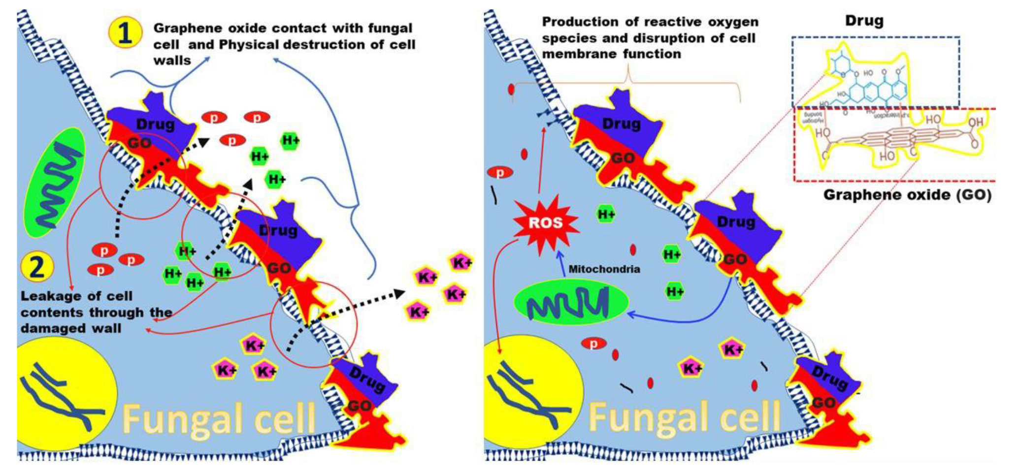
Figure 1. Functions and mechanisms of graphene oxide nanoparticles against fungi.
Cyto and genotoxicity of nano-graphene oxide
Suitability of pulmonary-targeted delivery using graphene oxide is determined by their increased efficacy, stability, and, most importantly, its toxicity and safety profile. Nano-graphene oxide can be considered an acceptable example of a safe design. Despite the potential benefits, the toxicity associated with inhalation exposure to this nanomaterial has not been fully elucidated. Towards this goal, Han et al. reported that the acute inhalation toxicity associated with NGO was low, while the In Vivo animal model studies demonstrated potential reproductive genotoxicities [ 99 , 100 ].
In a recent study, the NGO was administered via inhalation to male rats to examine their short-term In Vivo biodistribution [ 72 ]. According to their results, no remarkable systemic toxic effects were observed in the bronchoalveolar lavage cells, including the lymphocytes and macrophages. The results also exhibited a similar trend to those of a previous NGO exposure study [ 101 ]. Contrary to these results, another study using nano-graphene with sizes of <2, 5, and 20 µm administered via pharyngeal aspiration, reported increased inflammatory markers in the bronchoalveolar lavage fluids [ 102 ]. Such differing outcomes may be attributed to the varying size distributions of graphite nanoplates used across the studies. The NGO with a lateral size of 200-500 nm has been shown to induce cyto-genotoxic effects in human lung fibroblasts in a dose-dependent manner (1-100 μg/ml) via the mechanism of oxidative stress and the surface charge properties of NGO [ 103 ]. On the cellular level, an NGO with a lateral size range of 160-780 nm did not penetrate A549 cells but did induce oxidative stress in a dose-dependent manner and resulted in a slight decrease in cell viability at high concentrations, suggesting that NGO may be a safe material [ 104 ]. Additionally, the choice of delivery methods, such as pharyngeal aspiration techniques or inhalation of aerosolized materials, can also influence the inflammatory response. When pharyngeal aspiration and/or intratracheal instillation techniques are used to administer nano-carriers for drug delivery to the lungs, it should be considered that they can lead to increased aggregation of the nanostructures.
In a preclinical study, the intratracheal instillation of NGO in male Sprague-Dawley rats using a nose-only inhalation exposure system showed no significant inflammatory response [ 72 ]. In contrast, another study reported an increased inflammatory response, including an elevated acute phase response and elevated serum amyloid A levels, in mice following the intratracheal instillation of NGO and reduced graphene [ 105 ].
In a study, the genotoxicity of reduced graphene oxide nanoribbons in human mesenchymal stem cell lines was measured to assess the extent of DNA damage. According to their results, nanoparticles demonstrated genotoxicity at a 1.0 μg/mL concentration with DNA fragmentations and chromosomal aberrations [ 106 ]. Moreover, pristine NGO cytotoxicity toward bone marrow-derived mesenchymal stem cells was reported to be concentration-dependent [ 107 ]. These findings confirm the important role that the shape and dispersion status of graphene nanomaterials play in their interaction with cell lines and the subsequent induction of toxicity.
Biocompatibility of nano-graphene oxide
The NGO can be loaded into a kind of biodegradable polymeric composite to improve its physicomechanical properties [ 108 ]. Although these polymer-hybrid graphene materials are utilized in antimicrobial applications, they are not expected to compromise biocompatibility; rather, they are expected to facilitate the reduction and suppression of microbial growth [ 109 ]. Several studies have reported a decrease in fungal viability when exposed to NGO in nanocomposites [ 110 ]. Various physicochemical characteristics of NGO, including ROS, layer number and thickness, and sharp edges, all contribute to this decline.
Furthermore, the concentration of NGO and the exposure time can enhance the antifungal activity and negatively affect biodegradability and biocompatibility.
Various In Vivo and in vitro studies have been used to understand the biocompatibility of NGO-based compounds. In one study, poly (acrylic acid) and poly (ethylene glycol) were used to cover the NGO at different doses. The results obtained in their study indicated the extraordinary biocompatibility of poly (acrylic acid), compared to PEGylation for the functionalization of NGO [ 111 ].
In another study, NGO-modified chitosan was synthesized, and its antifungal activity against Fusarium graminearum was investigated [ 112 ]. In vitro studies reported that the combination of NGO and chitosan exhibited a notable synergistic inhibitory effect on the mycelial growth of F. graminearum, compared to a single NGO or chitosan. According to In Vivo studies, the NGO-chitosan composite demonstrated a significant reduction in both the incidence and severity of the disease, compared to a single NGO or chitosan, and the control efficacy of the NGO-chitosan composite reached 60.01%. In an unprecedented study, for the first time, the fungicidal activity of hybrid nano-compounds based on silver/reduced GO )rGO/Ag( was tested against clinical isolates of Candida spp. [ 68 ]. The antifungal susceptibility assay was conducted using the microdilution procedure based on modified guidelines from the Clinical and Laboratory Standards Institute. The highest inhibitory activity against Candida species was observed in rGO/Ag12% at a minimum of 6 h. However, after 2 h, initial antifungal interference was observed. None of the hybrid formulations of rGO/Ag nanocomposites exhibited toxicity towards G. mellonella. The conclusions are of paramount importance for clinical studies. Candida albicans is the predominant pathogenic fungus frequently isolated in nosocomial infections among immunocompromised patients [ 113 ]. The ability of this fungus to convert from yeast to hyphal morphology plays an important role in the formation of resistant biofilms, which effectively protect C. albicans against the effects of antifungal agents [ 114 ]. Recently, a study was conducted to investigate the effectiveness of zinc oxide nanorods decorated on an NGO against the human pathogen C. albicans [ 93 ].
The results obtained in their study demonstrated a significant reduction in the viability of C. albicans yeasts and an impact on hyphal development. The toxicity of NGO-Zinc oxide nanorods in HaCaT human keratinocyte cells showed that the nanomaterial was not toxic when glycerol was added as an emulsifier to the suspensions. The outcomes illustrated that NGO-Zinc oxide induced mortality in yeast cells via ROS formation, indicating its potential as an antifungal nanomaterial with extraordinary biocompatibility on human cells.
In another study, the synergistic antifungal potential of NGO-ethylene diamine tetraacetic acid (EDTA) and NGO-chitosan was evaluated against C. albicans [ 74 ]. The in vitro zone of inhibition outcomes verified that the nanocomposites showed inhibitory activity against C. albicans (16.0 mm for NGO-chitosan and 22.0 mm for NGO-EDTA). Notably, fungal cells may ultimately be damaged when they interact with NGO composites due to several mechanisms, including oxidative stress, wrapping/isolation, and membrane stress.
A novel approach for antifungal activities is the reaction of biocompatible NGO composite with amino acids, including Methionine and Cysteine in the presence of cellulose [ 115 ]. The method used for the formation of these antimicrobial nanocomposites involved two steps: the first step was the periodate oxidation of cellulose, and the second step was the reaction with amino acids in the presence of NGO. Both dialdehyde cellulose/NGO/Met and dialdehyde cellulose/NGO/Cys exhibited promising antifungal activity against the unicellular fungi C. albicans and Cryptococcus neoformans.
In an exciting study, a ternary NGO composite was prepared using chitosan and cerium oxide as a therapeutic platform against fungal and bacterial infections as well as breast cancer [ 116 ]. The results indicated the effective therapeutic behavior of the NGO/chitosan/cerium oxide composite against Aspergillus niger, Escherichia coli, Staphylococcus aureus, and Salmonella species. Furthermore, the NGO/chitosan/cerium composite exhibited outstanding anti-cancer potential on the MCF-7 cell line, with only 18.8% of cells remaining viable when exposed to the maximum concentration of 1,000 µg/mL of the nanocomposite. In addition, cerium oxide incorporation with chitosan enhanced their interaction and their reactivity.
Antifungal drugs delivery by nano-graphene oxide
The NGO is a material with amphiphilic properties, possessing a unique high surface area, which makes it a promising functional excipient for efficient drug loading and targeting. Recently, a study was conducted to investigate the antifungal activities and clotrimazole drug release profiles of NGOs incorporated into a buccal polyelectrolyte film [ 117 ]. The mucoadhesive excipients alginate and chitosan were chosen to create a polyelectrolyte film incorporating clotrimazole. According to their findings, the inclusion of NGO in the formulations resulted in a significant release of clotrimazole from the buccal films at pH 6.8, which could potentially be attributed to NGO modifying the electrostatic interactions between alginate and chitosan. The researchers have claimed that the antifungal activity against C. albicans was significantly higher in the formulation containing 0.04 wt% NGO, compared to the formulation without NGO. Nonetheless, when the NGO concentration increased to 0.09 wt%, the antifungal efficacy of the films was similar to that of the films with 0.04 wt% NGO, implying that the hydrophobic and electrostatic interactions between clotrimazole and NGO may also modulate the antifungal activity of clotrimazole. A similar study employed 1 wt% NGO in the formulations to evaluate the effect of NGO on the release and antifungal activity of clotrimazole from chitosan/alginate-based foams in vitro and affirmed the lower amount of NGO for enhancing the antifungal effect [ 95 ].
Recently, a novel formulation of NGO/fluconazole has been developed to examine its antifungal activity against fluconazole-resistant C. albicans [ 94 ]. The results indicated that the NGO/fluconazole formulation with minimal cytotoxic effect on the SW480 cell line was suitable for drug release in the Flui phosphate-buffered saline medium (72.42%). Notably, this formulation could enhance the antifungal effect against fluconazole-resistant C. albicans through DNA fragmentation, compared to NGO and fluconazole. Other studies have shown that incorporating fluconazole with different drug delivery systems, such as PEG-gold nanorods and cholesterol-gold nanorods, enhances the anti-mycotic effect of the drug against various strains of C. albicans [ 118 ]. However, the loading efficiency percentage of fluconazole on the NGO was higher (72.42%), compared to PEG-gold nanorods (62.5%) and cholesterol-gold nanorods (33.0%). Cholesterol/fluconazole/gold nanorods exhibited higher antifungal activity with a MIC of 0.25 nM, compared to that of PEG/fluconazole/gold nanorods (with MIC of 0.125 nM). The observed difference in antifungal activity can be attributed to the pattern of fluconazole release from formulations.
In a study, in vitro and In Vivo experiments were carried out to investigate whether NGO-composite containing indolicidin inhibited C. albicans yeasts [ 92 ]. This formulation exhibited favorable antifungal properties (MIC=3.12 μg/mL), compared to indolicidin (MIC 12.5 μg/mL) and fluconazole (MIC 4 μg/mL) in the cell line model. In In Vivo experiments, the NGO-indolicidin formulation eliminated the C. albicans infection in the liver and spleen of BALB/c mice, similar to fluconazole (p=0.001). Notably, incorporating indolicidin with gold nanoparticles appeared to reduce the antifungal activity of indolicidin, compared to the NGO-containing indolicidin formulation against fluconazole-resistant strains of C. albicans (MIC≥4) [ 119 ]. In contrast, no significant inhibitory effect of this formulation was observed, compared to fluconazole and amphotericin B on standard strains of Aspergillus spp. [ 120 ]. These outcomes are consistent with the fact that the integrity of the Aspergillus cell wall can prevent nanocomposite penetration. In a preliminary study, an in vitro experiment was carried out to investigate whether Amphotericin-B-loaded polymer-NGO was effective against Leishmania amazonensis cells after chemo-photothermal therapy [ 121 ].
Antiproliferative effects of the NGO-polyethyleneimine-Amphotericin-B formulation exhibited superior efficacy against the parasitic cells, compared to NGO-polyethyleneimine, which this activity was further potentiated under near-infrared (NIR) light irradiation. The scanning-transmission electron microscopy analysis of L. amazonensis promastigotes cells provided evidence indicative of the induction of autophagic and necrotic pathways leading to cellular demise. In contrast, no significant differences were observed in drug release from the NGO-Pluronic® P123- P123-Amphotericin-B formulation in the presence and absence of NIR light. In addition, cytotoxicity studies showed that the NGO-polyethyleneimine-Amphotericin-B formulation was nontoxic to mammalian host macrophages, whereas rGO-Pluronic® P123-Amphotericin-B was very toxic. Therefore, the facile synthesis, excellent photothermal effect, and extraordinary loading capacity make the NGO platform a promising Candidate for topical therapy.
Remarks and conclusions
To date, considerable research has been conducted to develop targeted therapies for the effective treatment of fungal diseases. Fungal infections present significant risks to human health, particularly for immunocompromised patients [ 122 , 123 ]. Furthermore, the increasing resistance of fungi to conventional antifungal agents heightens the urgent need for innovative treatment approaches. Understanding the biology of fungi is very important in order to identify therapeutic targets, which include exploring unique cellular pathways and structures that are present in fungi but not in human cells [ 124 , 125 ]. The main challenge is to develop drug delivery systems with maximum efficacy on fungal cells while minimizing toxicity on human cells; therefore, the use of medical nanotechnology is a suitable option. By using its products, it is possible to increase the effectiveness of the treatment while concurrently reducing side effects through the direct and targeted delivery of antifungal agents to the site of infection [ 126 , 127 ].
Insufficient delivery of the drug to the site of infection, where colonies of fungal cells remain intact, may inevitably lead to incomplete eradication of all unhealthy cells and result in the failure of the treatment modality, as well as further invasion and relapse of the disease [ 128 ]. The homogeneous diffusion of drug molecules within the site of infection is an essential step that is made possible by overcoming the integrity of the fungal cell wall [ 129 ]. Given that the major purpose of a fungal therapy strategy is to enhance the effectiveness of treatment modalities, a synergistic combination of drug delivery can be considered a vigorous approach that may guarantee the success of therapy [ 130 ].
Therefore, the appropriate response of these nanosystems to specific fungal stimuli, such as pH changes or special enzymes, requires proper engineering [ 131 ]. This engineering involves a multidisciplinary approach, including molecular biology, medical mycology, and pharmacology, to improve the ability to transport and protect controlled antifungal agents, increase treatment efficacy, overcome drug resistance, and ultimately minimize the risk of toxicity associated with high doses of drugs to achieve successful outcomes [ 132 ].
Although one of the main problems associated with currently used antifungal treatments is the inevitable emergence of adverse reactions, targeted drug delivery may potentially resolve the issue of side effects to some extent [ 133 ]. The NGOs appear to be promising nanocarriers for antimicrobial drugs due to their specific characteristics that contribute to the synergistic effect [ 126 ].
Conclusion
In conclusion, the utilization of an NGO as an innovative drug carrier presents a promising outlook for the treatment of pulmonary fungal infections. The unique physicochemical properties of this material, such as the generation of ROS, disruption of fungal cell membranes, and the capability to load antifungal drugs at lower doses, can enhance therapeutic efficacy while reducing side effects. However, challenges, such as potential toxicity, the need for surface modifications to improve biocompatibility, and an incomplete understanding of its mechanisms of action in the respiratory system warrant further investigation.
Overall, NGO, due to its potent antifungal properties, ability to deliver targeted drugs, and potential to mitigate drug resistance, is considered a promising Candidate for treating drug-resistant fungal infections. Nevertheless, comprehensive research on the design of drug delivery systems and the evaluation of biocompatibility under clinical conditions is essential to ensure the safe and effective application of this technology.
Acknowledgments
The authors would like to express their gratitude to the team of doctors and staff of the Anesthesiology Research Center, Department of Anesthesiology, Alzahra Hospital, Guilan University of Medical Sciences, Guilan, Iran for their assistance in the conduction of this study.
Conflicts of interest
The authors declare that they have no conflict of interest regarding the publication of this article.
Authors’ contribution
N. R. designed the study, and N. R., A. R., and A. H. collected and analyzed the data. N. R., I. H., and A. R. contributed to the first draft preparation and data collection. N. R. wrote the manuscript. I. H., A. D-M., and SH. Y-CH. improved and edited the manuscript. All authors read and confirmed the final version of the manuscript for publication.
Financial disclosure
No financial interests related to the material of this manuscript have been declared.
References
- Brackin AP, Hemmings SJ, Fisher MC, Rhodes J. Fungal genomics in respiratory medicine: what, how and when? Mycopathologia. 2021; 186(5):589-608.
- Collaborators GBDURIOM. Global, regional, and national burden of upper respiratory infections and otitis media, 1990-2021: a systematic analysis from the Global Burden of Disease Study 2021. Lancet Infect Dis. 2025; 25(1):36-51.
- Vuong NN, Hammond D, Kontoyiannis DP. Clinical Uses of Inhaled Antifungals for Invasive Pulmonary Fungal Disease: Promises and Challenges. J Fungi (Basel). 2023; 9(4):464.
- Foppiano Palacios C, Spichler Moffarah A. Diagnosis of pneumonia due to invasive molds. Diagnostics. 2021; 11(7):1226.
- Lass-Flörl C, Steixner S. The changing epidemiology of fungal infections. Mol Aspects Med. 2023; 94:101215.
- Hedayati MT, Khodavaisy S, Aliali M. A review on invasive aspergillosis in patients admitted to intensive care unit with emphasis on diagnostic methods. J Mazandaran Univ Med Sci. 2010; 19(74):99-112.
- Nguyen HN, Chaves-Lopez C, Oliveira RC, Paparella A, Rodrigues DF. Cellular and metabolic approaches to investigate the effects of graphene and graphene oxide in the fungi Aspergillus flavus and Aspergillus niger. Carbon. 2019; 143:419-29.
- Zhang W, Zhang H, Zhan M, Jing R, Wang X, Zhang Z. Epidemiological characteristics of invasive Aspergillus isolates: Morphology, drug susceptibility, and mutations in azole drug targets. J Infect Public Health. 2025; 18(1):102612.
- Fukuda Y, Homma T, Suzuki S, Takuma T, Tanaka A, Yokoe T, et al. High burden of Aspergillusfumigatus infection among chronic respiratory diseases. Chron Respir Dis. 2018; 15(3):279-85.
- Moore-Landecker E. Fungal spores. eLS. 2011.
- Kendrick B. Fungi: ecological importance and impact on humans. eLS. 2011.
- Koulenti D, Arvaniti K, Judd M, Lalos N, Tjoeng I, Xu E, et al. Ventilator-Associated Tracheobronchitis: To Treat or Not to Treat? Antibiotics (Basel). 2020; 9(2):51.
- Melsen WG, Rovers MM, Groenwold RH, Bergmans DC, Camus C, Bauer TT, et al. Attributable mortality of ventilator-associated pneumonia: a meta-analysis of individual patient data from randomised prevention studies. Lancet Infect Dis. 2013; 13(8):665-71.
- Klompas M, Branson R, Cawcutt K, Crist M, Eichenwald EC, Greene LR, et al. Strategies to prevent ventilator-associated pneumonia, ventilator-associated events, and nonventilator hospital-acquired pneumonia in acute-care hospitals: 2022 Update. Infect Control Hosp Epidemiol. 2022; 43(6):687-713.
- Forel JM, Voillet F, Pulina D, Gacouin A, Perrin G, Barrau K, et al. Ventilator-associated pneumonia and ICU mortality in severe ARDS patients ventilated according to a lung-protective strategy. Crit Care. 2012; 16(2):R65.
- Maheswary D, Leela KV, Ravi S. Perspectives of Novel Drug Delivery in Mycoses. Adv Nov Formul Drug Deliv. 2023;197-205.
- Kumar M, Hilles AR, Almurisi SH, Bhatia A, Mahmood S. Micro and nano-carriers-based pulmonary drug delivery system: Their current updates, challenges, and limitations–A review. JCIS Open. 2023; 12:100095.
- de Almeida Campos L, Fin MT, Santos KS, de Lima Gualque MW, Freire Cabral AKL, Khalil NM, et al. Nanotechnology-Based Approaches for Voriconazole Delivery Applied to Invasive Fungal Infections. Pharmaceutics. 2023; 15(1):266.
- Patel M, Mazumder R, Mishra R, Kant Kaushik K. Potential of Nanotechnology-based Formulations in Combating Pulmonary Infectious Diseases: A Current Scenario. Curr Pharm Des. 2022; 28(42):3413-27.
- Sunder S, Bhandari K, Sounkaria S, Vyas M, Singh BP, Chandra P. Antibiotics and nano-antibiotics in treatment of lung infection: In management of COVID-19. Microb Pathog. 2023; 184:106356.
- He Y, Zhang W, Xiao Q, Fan L, Huang D, Chen W, et al. Liposomes and liposome-like nanoparticles: From anti-fungal infection to the COVID-19 pandemic treatment. Asian J Pharm Sci. 2022; 17(6):817-37.
- Cheng SN, Tan ZG, Pandey M, Srichana T, Pichika MR, Gorain B, et al. A Critical Review on Emerging Trends in Dry Powder Inhaler Formulation for the Treatment of Pulmonary Aspergillosis. Pharmaceutics. 2020; 12(12):1161.
- Waghule T, Sankar S, Rapalli VK, Gorantla S, Dubey SK, Chellappan DK, et al. Emerging role of nanocarriers based topical delivery of anti-fungal agents in combating growing fungal infections. Dermatol Ther. 2020; 33(6):e13905.
- Marx-Stoelting P, Knebel C, Braeuning A. The Connection of Azole Fungicides with Xeno-Sensing Nuclear Receptors, Drug Metabolism and Hepatotoxicity. Cells. 2020; 9(5):1192.
- Lu H, Zhang S, Wang J, Chen Q. A Review on Polymer and Lipid-Based Nanocarriers and Its Application to Nano-Pharmaceutical and Food-Based Systems. Front Nutr. 2021; 8:783831.
- Conte G, Costabile G, Baldassi D, Rondelli V, Bassi R, Colombo D, et al. Hybrid lipid/polymer nanoparticles to tackle the cystic fibrosis mucus barrier in siRNA delivery to the lungs: does PEGylation make the difference? ACS Applied Materials & Interfaces. 2022; 14(6):7565-78.
- Adler-Moore J, Proffitt RT. AmBisome: liposomal formulation, structure, mechanism of action and pre-clinical experience. J Antimicrob Chemother. 2002; 49 Suppl 1(suppl_1):21-30.
- Seify R, Zahednezhad F, Zakeri-Milani P, Valizadeh H. Amphotericin B liposomal formulation: applicable preparation methods, challenges, and tips. Drug Dev Ind Pharm. 2023; 49(5):367-76.
- Connors KA, Amidon GL, Stella VJ. Chemical stability of pharmaceuticals: a handbook for pharmacists: John Wiley & Sons; 1986.
- Moen MD, Lyseng-Williamson KA, Scott LJ. Liposomal amphotericin B: a review of its use as empirical therapy in febrile neutropenia and in the treatment of invasive fungal infections. Drugs. 2009; 69:361-92.
- Daniyal M, Liu B, Wang W. Comprehensive Review on Graphene Oxide for Use in Drug Delivery System. Curr Med Chem. 2020; 27(22):3665-85.
- Liu J, Cui L, Losic D. Graphene and graphene oxide as new nanocarriers for drug delivery applications. Acta biomaterialia. 2013; 9(12):9243-57.
- Zare P, Aleemardani M, Seifalian A, Bagher Z, Seifalian AM. Graphene Oxide: Opportunities and Challenges in Biomedicine. Nanomaterials (Basel). 2021; 11(5):1083.
- Stone NR, Bicanic T, Salim R, Hope W. Liposomal amphotericin B (AmBisome®): a review of the pharmacokinetics, pharmacodynamics, clinical experience and future directions. Drugs. 2016; 76:485-500.
- LaMastro V, Campbell KM, Gonzalez P, Meng-Saccoccio T, Shukla A. Antifungal liposomes: Lipid saturation and cholesterol concentration impact interaction with fungal and mammalian cells. J Biomed Mater Res A. 2023; 111(5):644-59.
- Samad A, Sultana Y, Aqil M. Liposomal drug delivery systems: an update review. Curr Drug Deliv. 2007; 4(4):297-305.
- Alghuthaymi MA, C R, P R, Kalia A, Bhardwaj K, Bhardwaj P, et al. Nanohybrid Antifungals for Control of Plant Diseases: Current Status and Future Perspectives. J Fungi (Basel). 2021; 7(1):48.
- Zhang RX, Ahmed T, Li LY, Li J, Abbasi AZ, Wu XY. Design of nanocarriers for nanoscale drug delivery to enhance cancer treatment using hybrid polymer and lipid building blocks. Nanoscale. 2017; 9(4):1334-55.
- Tarannum N, Pooja K, Jakhar S, Mavi A. Nanoparticles assisted intra and transdermic delivery of antifungal ointment: an updated review. Discov Nano. 2024; 19(1)
- Leon-Buitimea A, Garza-Cervantes JA, Gallegos-Alvarado DY, Osorio-Concepcion M, Morones-Ramirez JR. Nanomaterial-Based Antifungal Therapies to Combat Fungal Diseases Aspergillosis, Coccidioidomycosis, Mucormycosis, and Candidiasis. Pathogens. 2021; 10(10):1303.
- Slavin YN, Bach H. Mechanisms of Antifungal Properties of Metal Nanoparticles. Nanomaterials (Basel). 2022; 12(24):4470.
- Carmo P, Garcia MT, Figueiredo-Godoi LMA, Lage ACP, Silva NSD, Junqueira JC. Metal Nanoparticles to Combat Candida albicans Infections: An Update. Microorganisms. 2023; 11(1):138.
- Silva LN, de Mello TP, de Souza Ramos L, Branquinha MH, Dos Santos ALS. Current Challenges and Updates on the Therapy of Fungal Infections. Curr Top Med Chem. 2019; 19(7):495-9.
- Osman N, Kaneko K, Carini V, Saleem I. Carriers for the targeted delivery of aerosolized macromolecules for pulmonary pathologies. Expert Opin Drug Deliv. 2018; 15(8):821-34.
- Merchant Z, Buckton G, Taylor KM, Stapleton P, Saleem IY, Zariwala MG, et al. A New Era of Pulmonary Delivery of Nano-antimicrobial Therapeutics to Treat Chronic Pulmonary Infections. Curr Pharm Des. 2016; 22(17):2577-98.
- Brunet K, Martellosio JP, Tewes F, Marchand S, Rammaert B. Inhaled Antifungal Agents for Treatment and Prophylaxis of Bronchopulmonary Invasive Mold Infections. Pharmaceutics. 2022; 14(3):641.
- Singh A, Sharma B, Mahto KK, Meis JF, Chowdhary A. High-Frequency Direct Detection of Triazole Resistance in Aspergillus fumigatus from Patients with Chronic Pulmonary Fungal Diseases in India. J Fungi (Basel). 2020; 6(2):67.
- Bassetti M, Vena A, Russo A, Peghin M. Inhaled Liposomal Antimicrobial Delivery in Lung Infections. Drugs. 2020; 80(13):1309-18.
- Wakaskar RR. General overview of lipid–polymer hybrid nanoparticles, dendrimers, micelles, liposomes, spongosomes and cubosomes. J Drug Target. 2018; 26(4):311-8.
- Westmeier D, Solouk-Saran D, Vallet C, Siemer S, Docter D, Gotz H, et al. Nanoparticle decoration impacts airborne fungal pathobiology. Proc Natl Acad Sci U S A. 2018; 115(27):7087-92.
- Napp J, Markus MA, Heck JG, Dullin C, Mobius W, Gorpas D, et al. Therapeutic Fluorescent Hybrid Nanoparticles for Traceable Delivery of Glucocorticoids to Inflammatory Sites. Theranostics. 2018; 8(22):6367-83.
- Kaur R, Dennison SR, Burrow AJ, Rudramurthy SM, Swami R, Gorki V, et al. Nebulised surface-active hybrid nanoparticles of voriconazole for pulmonary Aspergillosis demonstrate clathrin-mediated cellular uptake, improved antifungal efficacy and lung retention. J Nanobiotechnology. 2021; 19(1):19.
- Garcia-Mouton C, Hidalgo A, Cruz A, Pérez-Gil J. The Lord of the Lungs: The essential role of pulmonary surfactant upon inhalation of nanoparticles. Eur J Pharm Biopharm. 2019; 144:230-43.
- Pearson JP, Chater PI, Wilcox MD. The properties of the mucus barrier, a unique gel–how can nanoparticles cross it? Ther Deliv. 2016; 7(4):229-44.
- Hickey AJ, Mansour HM. Inhalation aerosols. physical and biological basis for therapy: CRC press; 2019.
- Aramendia I, Fernandez-Gamiz U, Lopez-Arraiza A, Rey-Santano C, Mielgo V, Basterretxea FJ, et al. Experimental Evaluation of Perfluorocarbon Aerosol Generation with Two Novel Nebulizer Prototypes. Pharmaceutics. 2019; 11(1):19.
- Matharu RK, Tabish TA, Trakoolwilaiwan T, Mansfield J, Moger J, Wu T, et al. Microstructure and antibacterial efficacy of graphene oxide nanocomposite fibres. J Colloid Interface Sci. 2020; 571:239-52.
- Seifi T, Kamali AR. Anti-pathogenic activity of graphene nanomaterials: A review. Colloids Surf B Biointerfaces. 2021; 199:111509.
- Rahmanian N, Eskandani M, Barar J, Omidi Y. Recent trends in targeted therapy of cancer using graphene oxide-modified multifunctional nanomedicines. J Drug Target. 2017; 25(3):202-15.
- Rahmanian N, Hamishehkar H, Dolatabadi JE, Arsalani N. Nano graphene oxide: a novel carrier for oral delivery of flavonoids. Colloids Surf B Biointerfaces. 2014; 123:331-8.
- Demirel E, Durmaz YY. PEGylated reduced graphene oxide as nanoplatform for targeted gene and drug delivery. Eur Polym J. 2023; 186:111841.
- Gungordu Er S, Edirisinghe M, Tabish TA. Graphene‐Based Nanocomposites as Antibacterial, Antiviral and Antifungal Agents. Adv Healthc Mater. 2023; 12(6):2201523.
- Wagner RD, Johnson SJ, Danielsen ZY, Lim JH, Mudalige T, Linder S. Polyethylene glycol-functionalized poly (Lactic Acid-co-Glycolic Acid) and graphene oxide nanoparticles induce pro-inflammatory and apoptotic responses in Candida albicans-infected vaginal epithelial cells. PLoS One. 2017; 12(4):e0175250.
- Diez-Orejas R, Feito MJ, Cicuendez M, Rojo JM, Portoles MT. Differential effects of graphene oxide nanosheets on Candida albicans phagocytosis by murine peritoneal macrophages. J Colloid Interface Sci. 2018; 512:665-73.
- Kahsay MH, Belachew N, Tadesse A, Basavaiah K. Magnetite nanoparticle decorated reduced graphene oxide for adsorptive removal of crystal violet and antifungal activities. RSC Adv. 2020; 10(57):34916-27.
- Wang X, Peng F, Cheng C, Chen L, Shi X, Gao X, et al. Synergistic Antifungal Activity of Graphene Oxide and Fungicides against Fusarium Head Blight In vitro and In Vivo. Nanomaterials (Basel). 2021; 11(9):2393.
- Zhang X, Cao H, Wang J, Li F, Zhao J. Graphene Oxide Exhibits Antifungal Activity against Bipolaris sorokiniana In vitro and In Vivo. Microorganisms. 2022; 10(10):1994.
- Gottardo B, Zoccal ARM, Maschio-Lima T, Lemes TH, Paziani MH, Von Zeska Kress MR, et al. Antifungal Activity of Nontoxic Nanocomposite Based on Silver and Reduced Graphene Oxide against Dermatophytes and Candida spp. ACS Biomater Sci Eng. 2023; 9(12):6870-9.
- Selvi MSG, Jauhar RM, Sivaraj D, Suvitha A, Malarvizhi V, Vijayaraghavan G. Enhanced antifungal properties of the Ag-decorated GO and rGO nanocomposites for medical applications. Bull Mater Sci. 2024; 47(1):27.
- Wang X, Cai A, Wen X, Jing D, Qi H, Yuan H. Graphene oxide-Fe3O4 nanocomposites as high-performance antifungal agents against Plasmopara viticola. Sci China Mater. 2017; 60(3):258-68.
- Wang X, Liu C, Li H, Zhang H, Ma R, Zhang Q, et al. Metabonomics-assisted label-free quantitative proteomic and transcriptomic analysis reveals novel insights into the antifungal effect of graphene oxide for controlling Fusarium graminearum. Environ Sci Nano. 2019; 6(11):3401-21.
- Kim YH, Jo MS, Kim JK, Shin JH, Baek JE, Park HS, et al. Short-term inhalation study of graphene oxide nanoplates. Nanotoxicology. 2018; 12(3):224-38.
- Li C, Wang X, Chen F, Zhang C, Zhi X, Wang K, et al. The antifungal activity of graphene oxide-silver nanocomposites. Biomaterials. 2013; 34(15):3882-90.
- Khalil WF, El-Sayyad GS, El Rouby WM, Sadek M, Farghali AA, El-Batal AI. Graphene oxide-based nanocomposites (GO-chitosan and GO-EDTA) for outstanding antimicrobial potential against some Candida species and pathogenic bacteria. Int J Biol Macromol. 2020; 164:1370-83.
- Pham VT, Truong VK, Quinn MD, Notley SM, Guo Y, Baulin VA, et al. Graphene induces formation of pores that kill spherical and rod-shaped bacteria. ACS nano. 2015; 9(8):8458-67.
- Ou L, Song B, Liang H, Liu J, Feng X, Deng B, et al. Toxicity of graphene-family nanoparticles: a general review of the origins and mechanisms. Part Fibre Toxicol. 2016; 13(1):57.
- Li C, Lu Y, Yan J, Yu W, Zhao R, Du S, et al. Effect of long-term ageing on graphene oxide: structure and thermal decomposition. R Soc Open Sci. 2021; 8(12):202309.
- Li R, Liu C, Wan C, Liu T, Zhang R, Du J, et al. A Targeted and pH-Responsive Nano-Graphene Oxide Nanoparticle Loaded with Doxorubicin for Synergetic Chemo-Photothermal Therapy of Oral Squamous Cell Carcinoma. Int J Nanomedicine. 2023; 18:3309-24.
- Kumbhar P, Kaur J, De Rubis G, Paudel KR, Prasher P, Patel VK, et al. Inhalation drug delivery in combating pulmonary infections: Advances and challenges. J Drug Deliv Sci Technol. 2023; 89:105022.
- Rayannavar S, Shaha B, Shankargouda S, Nelogi S. Antifungal efficacy of tissue conditioner incorporated with nano graphene oxide: An in-vitro study. IJDSIR. 2020; 3:601-7.
- Jasim DA, Menard-Moyon C, Begin D, Bianco A, Kostarelos K. Tissue distribution and urinary excretion of intravenously administered chemically functionalized graphene oxide sheets. Chem Sci. 2015; 6(7):3952-64.
- Lehner BAE, Benz D, Moshkalev SA, Meyer AS, Cotta MA, Janissen R. Biocompatible Graphene Oxide Nanosheets Densely Functionalized with Biologically Active Molecules for Biosensing Applications. ACS Appl Nano Mater. 2021; 4(8):8334-42.
- AbouAitah K, Sabbagh F, Kim BS. Graphene oxide nanostructures as nanoplatforms for delivering natural therapeutic agents: Applications in cancer treatment, bacterial infections, and bone regeneration medicine. Nanomaterials. 2023; 13(19):2666.
- Lowe SE, Zhong YL. Challenges of Industrial‐Scale Graphene Oxide Production. Graphene oxide: fundamentals and applications. 2016;410-31.
- Zou X, Zhang L, Wang Z, Luo Y. Mechanisms of the Antimicrobial Activities of Graphene Materials. J Am Chem Soc. 2016; 138(7):2064-77.
- Mohammed H, Kumar A, Bekyarova E, Al-Hadeethi Y, Zhang X, Chen M, et al. Antimicrobial Mechanisms and Effectiveness of Graphene and Graphene-Functionalized Biomaterials. A Scope Review. Front Bioeng Biotechnol. 2020; 8:465.
- Yi X, Gao H. Cell interaction with graphene microsheets: near-orthogonal cutting versus parallel attachment. Nanoscale. 2015; 7(12):5457-67.
- He J, Zhu X, Qi Z, Wang C, Mao X, Zhu C, et al. Killing dental pathogens using antibacterial graphene oxide. ACS Appl Mater Interfaces. 2015; 7(9):5605-11.
- Whitehead K, Vaidya M, Liauw C, Brownson D, Ramalingam P, Kamieniak J, et al. Antimicrobial activity of graphene oxide-metal hybrids. Int Biodeterior Biodegradation. 2017; 123:182-90.
- Sandhya P, Jose J, Sreekala M, Padmanabhan M, Kalarikkal N, Thomas S. Reduced graphene oxide and ZnO decorated graphene for biomedical applications. Ceram Int. 2018; 44(13):15092-8.
- Higuchi Y. Chromosomal DNA fragmentation in apoptosis and necrosis induced by oxidative stress. Biochem Pharmacol. 2003; 66(8):1527-35.
- Farzanegan A, Roudbary M, Falahati M, Khoobi M, Gholibegloo E, Farahyar S, et al. Synthesis, characterization and antifungal activity of a novel formulated nanocomposite containing Indolicidin and Graphene oxide against disseminated candidiasis. J Mycol Med. 2018; 28(4):628-36.
- Ficociello G, De Caris MG, Trillo G, Cavallini D, Sarto MS, Uccelletti D, et al. Anti-Candidal Activity and In vitro Cytotoxicity Assessment of Graphene Nanoplatelets Decorated with Zinc Oxide Nanorods. Nanomaterials (Basel). 2018; 8(10):752.
- Asadi Shahi S, Roudbar Mohammadi S, Roudbary M, Delavari H. A new formulation of graphene oxide/fluconazole compound as a promising agent against Candida albicans. Prog Biomater. 2019; 8:43-50.
- Huang J, Jacobsen J, Genina N, Larsen SW, Nielsen HM, Mullertz A, et al. Investigating the effect of graphene oxide in chitosan/alginate-based foams on the release and antifungal activity of clotrimazole in vitro. Eur J Pharm Sci. 2022; 174:106204.
- Cobos M, De-La-Pinta I, Quindos G, Fernandez MJ, Fernandez MD. Graphene Oxide-Silver Nanoparticle Nanohybrids: Synthesis, Characterization, and Antimicrobial Properties. Nanomaterials (Basel). 2020; 10(2):376.
- Celebi K, Buchheim J, Wyss RM, Droudian A, Gasser P, Shorubalko I, et al. Ultimate permeation across atomically thin porous graphene. Science. 2014; 344(6181):289-92.
- Perreault F, de Faria AF, Nejati S, Elimelech M. Antimicrobial Properties of Graphene Oxide Nanosheets: Why Size Matters. ACS Nano. 2015; 9(7):7226-36.
- Han SG, Kim JK, Shin JH, Hwang JH, Lee JS, Kim TG, et al. Pulmonary Responses of Sprague-Dawley Rats in Single Inhalation Exposure to Graphene Oxide Nanomaterials. Biomed Res Int. 2015; 2015(1):376756.
- Ema M, Gamo M, Honda K. A review of toxicity studies on graphene-based nanomaterials in laboratory animals. Regul Toxicol Pharmacol. 2017; 85:7-24.
- Kim JK, Shin JH, Lee JS, Hwang JH, Lee JH, Baek JE, et al. 28-Day inhalation toxicity of graphene nanoplatelets in Sprague-Dawley rats. Nanotoxicology. 2016; 10(7):891-901.
- Roberts JR, Mercer RR, Stefaniak AB, Seehra MS, Geddam UK, Chaudhuri IS, et al. Evaluation of pulmonary and systemic toxicity following lung exposure to graphite nanoplates: a member of the graphene-based nanomaterial family. Part Fibre Toxicol. 2015; 13:1-22.
- Wang A, Pu K, Dong B, Liu Y, Zhang L, Zhang Z, et al. Role of surface charge and oxidative stress in cytotoxicity and genotoxicity of graphene oxide towards human lung fibroblast cells. J Appl Toxicol. 2013; 33(10):1156-64.
- Chang Y, Yang ST, Liu JH, Dong E, Wang Y, Cao A, et al. In vitro toxicity evaluation of graphene oxide on A549 cells. Toxicol Lett. 2011; 200(3):201-10.
- Bengtson S, Knudsen KB, Kyjovska ZO, Berthing T, Skaug V, Levin M, et al. Differences in inflammation and acute phase response but similar genotoxicity in mice following pulmonary exposure to graphene oxide and reduced graphene oxide. PLoS One. 2017; 12(6):e0178355.
- Akhavan O, Ghaderi E, Emamy H, Akhavan F. Genotoxicity of graphene nanoribbons in human mesenchymal stem cells. Carbon. 2013; 54:419-31.
- Zhang X, Wei C, Li Y, Li Y, Chen G, He Y, et al. Dose-dependent cytotoxicity induced by pristine graphene oxide nanosheets for potential bone tissue regeneration. J Biomed Mater Res A. 2020; 108(3):614-24.
- Lee SJ, Yoon SJ, Jeon IY. Graphene/Polymer Nanocomposites: Preparation, Mechanical Properties, and Application. Polymers (Basel). 2022; 14(21):4733.
- Gontarek-Castro E, Rybarczyk MK, Castro-Muñoz R, Morales-Jiménez M, Barragán-Huerta B, Lieder M. Characterization of PVDF/graphene nanocomposite membranes for water desalination with enhanced antifungal activity. Water. 2021; 13(9):1279.
- Alimardani V, Abolmaali SS, Borandeh S. Antifungal and antibacterial properties of graphene-based nanomaterials: a mini-review. J Nanostruct. 2019; 9(3):402-13.
- Xu M, Zhu J, Wang F, Xiong Y, Wu Y, Wang Q, et al. Improved in vitro and In Vivo biocompatibility of graphene oxide through surface modification: poly (acrylic acid)-functionalization is superior to PEGylation. ACS nano. 2016; 10(3):3267-81.
- Zhang W, Cheng C, Wang R, Peng F, Du H, Zheng Z, et al. Graphene oxide modification enhances the activity of chitosan against Fusarium graminearum in vitro and In Vivo. Int J Biol Macromol. 2022; 219:1112-21.
- Adiguzel N, Karakurt Z, Gungor G, Yazicioglu Mocin O, Acarturk E, Sogukpinar O, et al. Mortality rates and risk factors associated with nosocomial Candida infection in a respiratory intensive care unit. Tuberk Toraks. 2010; 58(1):35-43.
- Timsit JF, Schwebel C, Styfalova L, Cornet M, Poirier P, Forrestier C, et al. Impact of bronchial colonization with Candida spp. on the risk of bacterial ventilator-associated pneumonia in the ICU: the FUNGIBACT prospective cohort study. Intensive Care Med. 2019; 45(6):834-43.
- Hashem AH, Hasanin M, Kamel S, Dacrory S. A new approach for antimicrobial and antiviral activities of biocompatible nanocomposite based on cellulose, amino acid and graphene oxide. Colloids Surf B Biointerfaces. 2022; 209(Pt 1):112172.
- Saranya J, Preethi S, Shaik MR, Khan M, Khan M, Shaik B. Development of cerium oxide/chitosan/graphene oxide nanocomposite: an investigation toward its biological applications under in vitro conditions. Crystals. 2023; 13(9):1393.
- Huang J, Jacobsen J, Larsen S, Genina N, Van de Weert M, Müllertz A, et al. Graphene oxide as a functional excipient in buccal films for delivery of clotrimazole: Effect of molecular interactions on drug release and antifungal activity in vitro. Int J Pharm. 2020; 589:119811.
- Hamad KM, Mahmoud NN, Al-Dabash S, Al-Samad LA, Abdallah M, Al-Bakri AG. Fluconazole conjugated-gold nanorods as an antifungal nanomedicine with low cytotoxicity against human dermal fibroblasts. RSC advances. 2020; 10(43):25889-97.
- Rahimi H, Roudbarmohammadi S, Delavari HH, Roudbary M. Antifungal effects of indolicidin-conjugated gold nanoparticles against fluconazole-resistant strains of Candida albicans isolated from patients with burn infection. Int J Nanomedicine. 2019; 14:5323-38.
- Farzanegan A, Rudbary M, Falahati M. Evaluation of antifungal activity of graphene oxide conjugated with indolicidin against Aspergillus spp. and Candida albicans. Curr Med Mycol. 2016; 2(4)
- Vitorino LS, Dos Santos TC, Bessa IAA, Santos ECS, Vercoza BRF, de Oliveira LAS, et al. Amphotericin-B-loaded polymer-functionalized reduced graphene oxides for Leishmania amazonensis chemo-photothermal therapy. Colloids Surf B Biointerfaces. 2022; 209(Pt 1):112169.
- Little JS, Weiss ZF, Hammond SP. Invasive Fungal Infections and Targeted Therapies in Hematological Malignancies. J Fungi (Basel). 2021; 7(12):1058.
- Riley MM-S. Invasive fungal infections among immunocompromised patients in critical care settings: infection prevention risk mitigation. Crit Care Nurs Clin North Am. 2021; 33(4):395-405.
- Logan A, Wolfe A, Williamson JC. Antifungal Resistance and the Role of New Therapeutic Agents. Curr Infect Dis Rep. 2022; 24(9):105-16.
- Hasim S, Coleman JJ. Targeting the fungal cell wall: current therapies and implications for development of alternative antifungal agents. Future Med Chem. 2019; 11(8):869-83.
- Mali R, Patil J. Nanoparticles: A Novel Antifungal Drug Delivery System. Mater Proc. 2023; 14(1):61.
- Nami S, Aghebati-Maleki A, Aghebati-Maleki L. Current applications and prospects of nanoparticles for antifungal drug delivery. EXCLI J. 2021; 20:562-84.
- Cowen LE, Sanglard D, Howard SJ, Rogers PD, Perlin DS. Mechanisms of antifungal drug resistance. Cold Spring Harb Perspect Med. 2015; 5(7):a019752.
- Kang X, Kirui A, Muszynski A, Widanage MCD, Chen A, Azadi P, et al. Molecular architecture of fungal cell walls revealed by solid-state NMR. Nat Commun. 2018; 9(1):2747.
- Cui J, Ren B, Tong Y, Dai H, Zhang L. Synergistic combinations of antifungals and anti-virulence agents to fight against Candida albicans. Virulence. 2015; 6(4):362-71.
- Bernardos A, Bozik M, Montero A, Perez-Esteve E, Garcia-Casado E, Lhotka M, et al. Secreted Enzyme-Responsive System for Controlled Antifungal Agent Release. Nanomaterials (Basel). 2021; 11(5):1280.
- Mishra V, Singh M, Mishra Y, Charbe N, Nayak P, Sudhakar K, et al. Nanoarchitectures in management of fungal diseases: An overview. Applied Sciences. 2021; 11(15):7119.
- Horvat S, Yu Y, Manz H, Keller T, Beilhack A, Groll J, et al. Nanogels as Antifungal‐Drug Delivery System Against Aspergillus fumigatus. Adv NanoBiomed Res. 2021; 1(5):2000060.

