
Introduction
Chaetomium globosum is a rare causative agent of onychomycosis, typically found in soil, decaying plant material, and other organic matter. The genus Chaetomium is classified under the family Chaetomiaceae, class Sordariomycetes, and phylum Ascomycota [ 1 ]. It is a dematiaceous non-dermatophyte fungus, which is commonly found in the environment, but its involvement in human infections, particularly nail infections, is infrequent and often underreported. Its identification requires careful mycological workup, including direct microscopy and culture on appropriate media, followed by microscopic identification of characteristic perithecia and ascospores [ 2 ].
This case study aimed to report a case of onychomycosis in a patient who presented with brownish-yellow to black nail discoloration and subungual hyperkeratosis involving the right-hand nail. Direct microscopic examination of nail scrapings using potassium hydroxide preparation revealed septate hyphae. Cultures on Sabouraud's dextrose agar without cycloheximide consistently produced fast-growing colonies, initially appearing velvety white before turning dark grey to brown. Slide culture analysis revealed brown-colored septate hyphae, perithecia, and ascospores, confirming the presence of Chaetomium globosum. Species-level identification was performed by MALDI-TOF (Matrix-assisted laser desorption ionization–time of flight) mass spectrometry. This report contributed to increasing awareness about rare fungal etiologies in nail infections, particularly in patients exposed to soil and plant material.
Case Presentation
A 46-year-old male visited our department with progressive nail fragility and breakage of the right-hand nails over the past year. He presented with progressive nail discoloration. The discoloration began as a faint yellowish spot and gradually spread to involve the entire nail. He did not recall any trauma or any injury to nails or any other history of long-term administration of steroids or drugs. The patient had no known chronic illnesses, including diabetes, cardiovascular disease, or immunosuppressive conditions. He was a mason by occupation and a resident of West Champaran, Bihar, India.
On examination, the nail appeared thickened, brittle, and discolored with a yellowish-brown to black hue. The nail plate was dystrophic, with irregular ridging and subungual hyperkeratosis. There was no evidence of periungual inflammation or involvement of skin, including the scalp. General physical examination results were unremarkable (Figure 1A).
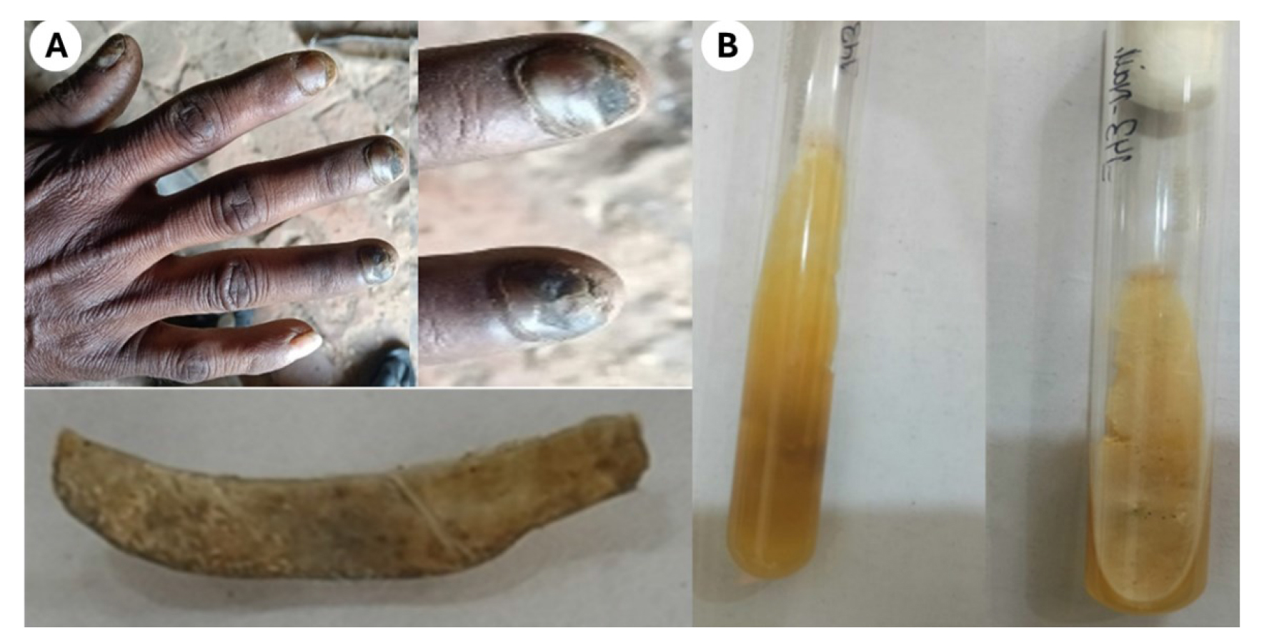
Figure 1.(A) Yellow brownish to black discoloration with hyperkeratosis on the right hand nails. Close-up view of the right-hand nails and Nail clipping, (B) Obverse and reverse surface of Sabouraud’s dextrose agar plate after incubation at 25 ᵒC for 5 days
Nail clippings were collected using a sterile nail cutter and subjected to 40% potassium hydroxide digestion. Direct microscopy revealed septate, branched hyphae.
Cultures on Sabouraud’s Dextrose Agar with chloramphenicol incubated at 25 °C showed rapid growth of a cottony white mycelial colony, which later turned grayish-green with dark speckles and a brown reverse. Lactophenol cotton blue mount revealed large, lemon-shaped perithecia with terminal setae, which is the characteristic of C. globosum (Figure 1B).
When fungal colonies were examined using slide cultures stained with lactophenol cotton blue, distinct morphological features were observed under light microscopy. The hyphae appeared brown and septate, while perithecia were large and dark brown to black, exhibiting a globose to flask-like shape with unbranched, hair-like filamentous appendages on their surface. These perithecia contained ostioles and enclosed asci, within which single-celled ascospores were present. The ascospores were olive brown in color and had a characteristic lemon-shaped appearance (Figures 2A and 2B).
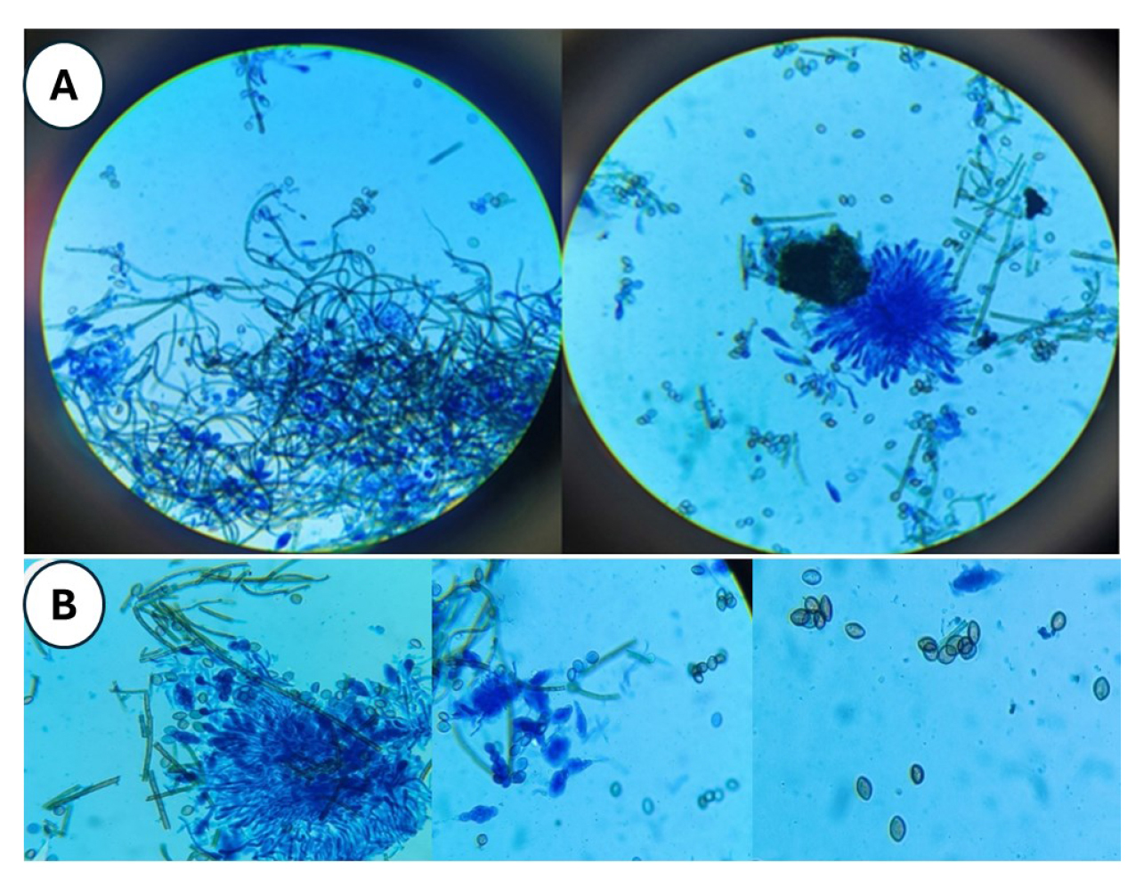
Figure 2.(A) Large, dark brown to black, flask-shaped perithecia with hair-like filamentous appendages (lactophenol cotton blue mount), (B) Close-up view of asci and single-celled ascospores: dark brown-colored septated hyphae and lemon-shaped ascospores (lactophenol cotton blue mount)
Antifungal susceptibility testing was performed using the broth microdilution method in accordance with Clinical and Laboratory Standards Institute (CLSI) M38: Reference Method for Broth Dilution Antifungal Susceptibility Testing of Filamentous Fungi [ 3 ]. The testing was conducted in RPMI 1640 medium. Itraconazole exhibited the highest in vitro efficacy with a minimum inhibitory concentration (MIC) of 0.25 µg/mL, followed by terbinafine (8 µg/mL), griseofulvin (16 µg/mL), and fluconazole, which showed the least activity (MIC: 32 µg/mL).
The patient was treated with oral itraconazole (200 mg/day for 12 weeks) plus topical 5% amorolfine nail lacquer twice weekly, resulting in marked clinical improvement with reduced discoloration and subungual hyperkeratosis (Figure 3).
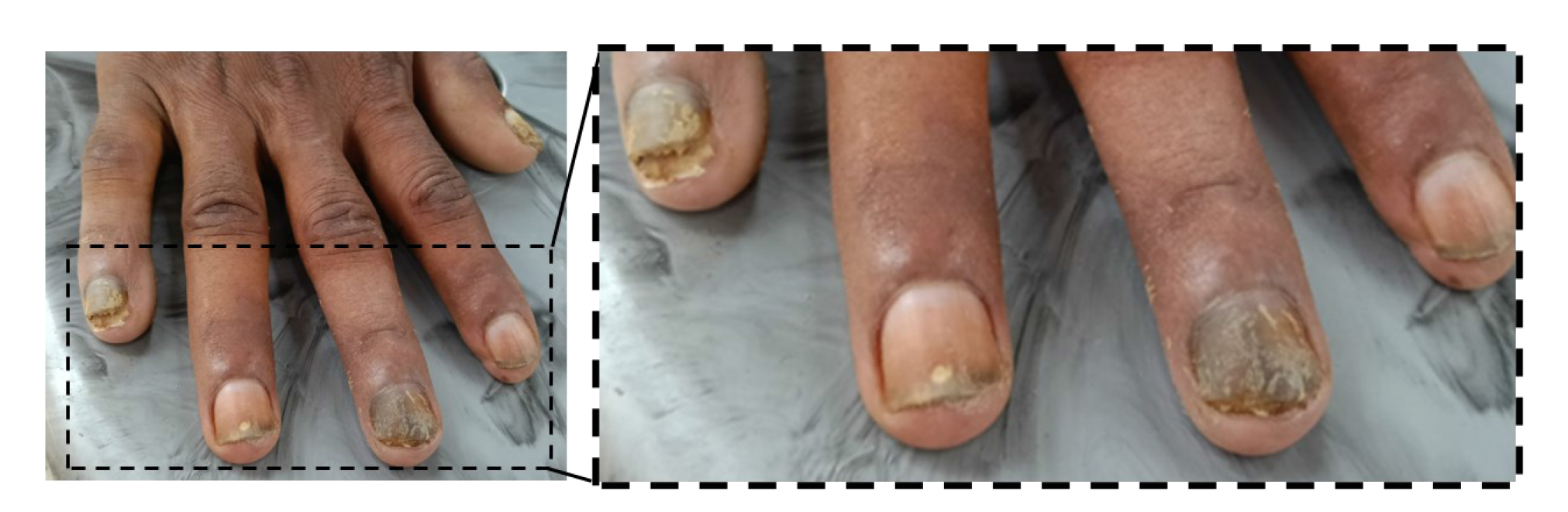
Figure 3. Follow-up photograph of the affected fingernail showing noticeable clinical improvement after treatment
Discussion
Chaetomium globosum is an uncommon cause of onychomycosis, primarily affecting immunocompromised individuals; however, cases in immunocompetent hosts have been reported. It is a saprophytic fungus commonly found in soil and decaying plant matter. Chaetomium is not commonly pathogenic; nevertheless, infections, such as cutaneous infections, onychomycosis, peritonitis, and brain abscesses have been reported [ 4 ]. The probable mode of infection in this case was traumatic inoculation while working in the fields.
Non-dermatophytic molds contribute to approximately 1.45-17.6% of cases [ 5 - 7 ]. Currently, the genus Chaetomium consists of 105 species since it was first described by Kunze in 1817. However, only a few species are recognized as human pathogens, including C. atrobrunneum, C. strumarium, C. perlucidum, C. funicolum, and C. globosum. Onychomycosis due to C. globosum is documented from India [ 8 , 9 ], Spain [ 10 ], Korea [ 1 ], Canada [ 11 , 12 ], and Japan [ 13 ].
The initial identification was based on morphological characteristics, revealing perithecia ranging from 175 to 280 µm in size, along with distinctive brown, septate setae. Chaetomium globosum was specifically identified by its brown, lemon-shaped ascospores measuring 9–12 × 7–9 µm and its inability to grow at a temperature of 42 °C. Clinical differentiation from dermatophytic onychomycosis is challenging, making mycological examination essential for accurate diagnosis [ 4 , 14 ]. MALDI-TOF provided reliable identification in the present case. However, sequence analysis of the internal transcribed spacer region of ribosomal DNA remains the gold standard for definitive species-level confirmation [ 15 ]. Nevertheless, it could not be performed due to resource limitations. This is acknowledged, and future studies should include this to improve the diagnosis of rare fungi.
Any disruption to the integrity of the corneal layer of the nail can allow fungi, including less pathogenic species, like Chaetomium spp., to penetrate more easily. Trauma is a key predisposing factor for onychomycosis and cutaneous lesions [ 16 ].
Additionally, occupation of an individual plays a crucial role in susceptibility to these infections. In the present case of nail infection, the patient was a mason, a profession that involves regular and prolonged exposure to materials, such as cement, sand, and soil. These substances are well-known reservoirs for various environmental fungi, including C. globosum. Additionally, the abrasive nature of cement may cause microabrasions to the nail bed and surrounding skin, allowing fungal entry and pathogenesis. Occupational exposure, along with trauma, likely contributed to the disruption of the corneal layer of the nail, facilitating fungal penetration.
In this case, antifungal testing showed itraconazole had the highest in vitro activity against C. globosum and was also effective clinically. These findings aligned with Guarro et al. [ 4 ], who found Chaetomium species susceptible to imidazoles, like itraconazole, while 5-fluorocytosine and amphotericin B were ineffective. Non-dermatophytic mold onychomycosis often responds poorly to standard antifungals [ 17 ]. Aspiroz et al. [ 10 ] achieved a complete cure with terbinafine; other studies support terbinafine and itraconazole efficacy. Similarly, in this case, oral itraconazole (200 mg/day) plus 5% amorolfine nail lacquer led to clinical improvement. However, complete clinical resolution could not be documented, as the patient did not return for extended follow-up after the initial improvement. The clinical outcome indicates that combining oral itraconazole with topical 5% amorolfine could be an effective treatment approach for rare non-dermatophytic fungi, like Chaetomium globosum.
Conclusion
Onychomycosis is a frequently occurring nail infection, primarily caused by dermatophytes. In some instances, non-dermatophytic molds may also be involved, though they are often overlooked due to limited awareness. However, if clinical findings and laboratory results confirm a fungal etiology, these molds should be recognized as pathogens and treated appropriately with antifungal therapy.
Acknowledgments
The authors are grateful to the Indian Council of Medical Research, Advanced Mycology Diagnostic and Research Centre, King George’s Medical University, Lucknow, India, for laboratory support and access to diagnostic facilities for this case study.
Conflicts of interest
The authors declare no conflict of interest.
Authors’ contributions
All authors contributed to case diagnosis, mycological workup, and drafting the manuscript.
Financial disclosure
No financial support was received for this study.
Ethics approval
Ethical approval was obtained from the Institutional Ethics Committee of King George's Medical University, Lucknow, India [Reference No: XXVI-PGTSC-IIA/P62].
References
- Kim DM, Lee MH, Suh MK, Ha GY, Kim H, Choi JS. Onychomycosis caused by Chaetomium globosum. Ann Dermatol. 2013; 25(2):232-6.
- Elewski BE. Onychomycosis: pathogenesis, diagnosis, and management. Clin Microbiol Rev. 1998; 11(3):415-29.
- Clinical and Laboratory Standards Institute (CLSI).. Reference method for broth dilution antifungal susceptibility testing of filamentous fungi. CLSI standard M38. Wayne, PA: CLSI; 2017.
- Guarro J, Soler L, Rinaldi MG. Pathogenicity and antifungal susceptibility of Chaetomium species. Eur J Clin Microbiol Infect Dis. 1995; 14(7):613-8.
- Cuomo CA, Untereiner WA, Ma LJ, Grabherr M, Birren BW. Draft genome sequence of the cellulolytic fungus Chaetomium globosum. Genome Announc. 2015; 3(1):e00021-15.
- Fernández MS, Rojas FD, Cattana ME, Sosa Mde L, Mangiaterra ML, Giusiano GE. Aspergillus terreus complex: an emergent opportunistic agent of Onychomycosis. Mycoses. 2013; 56(4): 477-81.
- Hwang SM, Suh MK, Ha GY. Onychomycosis due to nondermatophytic molds. Ann Dermatol. 2012; 24 (2): 175-80.
- Naidu J, Singh SM, Pouranik M. Onychomycosis caused by Chaetomium globosum Kunze. Mycopathologia. 1991; 113(1):31-4.
- Latha R, Sasikala R, Muruganandam N. Onychomycosis due to ascomycete Chaetomium globosum: A case report. Indian J Pathol Microbiol. 2010; 53(3):566-7.
- Aspiroz C, Gene J, Rezusta A. First Spanish case of onychomycosis caused by Chaetomium globosum. Med Mycol. 2007; 45(3):279-82.
- Jean L, Cellier E. A case report of a mixed Chaetomium globosum /Trichophyton mentagrophytes onychomycosis. Med Mycol Case Rep. 2012; 1:76-8.
- Stiller MJ, Rosenthal S, Summerbell RC, Pollack J, Chan A. Onychomycosis of the toenails caused by Chaetomium globosum. J Am Acad Dermatol. 1992; 26(5):775-6.
- Hattori N, Adachi M, Kaneko T, Shimozuma M, Ichinohe M, Iozumi K. Case report. Onychomycosis due to Chaetomium globosum successfully treated with itraconazole. Mycoses. 2000; 43(1-2):89-92.
- Abbott SP, Sigler L, McAleer R, McGough DA, Rinaldi MG, Mizell G. Fatal cerebral mycoses caused by the ascomycete Chaetomium strumarium. J Clin Microbiol. 1995; 33(10):2692-8.
- Yu J, Yang S, Zhao Y, Li R. A case of subcutaneous phaeohyphomycosis caused by Chaetomium globosum and the sequences analysis of C. globosum. Med Mycol. 2006; 44(6):541-5.
- Vander Straten MR, Balkis MM, Ghannoum MA. The role of nondermatophyte molds in onychomycosis: diagnosis and treatment. Dermatol Ther. 2002; 15(2):89-98.
- Nolting S, Brautigam M, Weidinger G. Terbinafine in onychomycosis with involvement by non‐dermatophytic fungi. Br J Dermatol. 1994; 130(s43):16-21.


