
Introduction
Otomycosis refers to a sub-acute or chronic inflammation and infection of the external auditory canal (EAC), which, if left untreated, can spread to the middle ear. Clinical hallmark symptoms that characterize this infection include itching, ear pain, otorrhea, pruritus, tinnitus, and hearing loss [ 1 , 2 ]. Despite the widespread distribution of otomycosis worldwide, it is more prevalent in regions with tropical and subtropical climates [ 3 ]. It is estimated to account for approximately 30% of all ear infections [ 4 ]. Otomycosis is more prevalent among individuals aged 21-40; however, conflicting results have been reported concerning its gender distribution [ 5 ]. In addition to climate characteristics, the risk factors contributing to this disease include swimming, use of hearing aids, long-term or excessive use of broad-spectrum antibiotics, anatomical abnormalities, immunodeficiency, and alterations of ear cerumen [ 6 , 7 ].
Although different species of yeasts and filamentous fungi can cause otomycosis, the dominant causative agents are Aspergillus species (i.e., Aspergillus section Nigri and Aspergillus section Flavi) and Candida species (i.e., C. albicans and C. parapsilosis). However, it is important to mention Candida auris as a multidrug-resistant emerging yeast. Since the first report of its involvement in the ear canal of a Japanese patient in 2009, C. auris has been isolated in over 50 countries across six continents. Its rapid transmissibility has resulted in many outbreaks worldwide, and it has been isolated from patients with otomycosis in several cases [ 8 - 11 ]. Unlikely, otomycoses rarely can develop due to dermatophytes or non-dermatophyte fungi, such as Penicillium species, Fusarium species, Mucoalean fungi, Scopulariopsis species, and Alternaria species [ 12 - 15 ]. Management and treatment of otomycosis are complicated due to the potential for recurrence of the infection and the development of drug resistance [ 4 , 16 ]. Identifying etiological agents is possible only through laboratory analyses, including mycological and bacteriological examination [ 17 ]. Effective and beneficial treatment of otomycosis involves removal of secretions and debris, and administration of appropriate local and, in rare cases, systemic antifungal agents [ 14 , 16 ], followed by management and control of predisposing factors. Although there are no universally approved treatment protocols and guidelines for otomycoses, it is worth noting that imidazole and triazole derivatives are the most commonly used antimycotic agents for local application [ 4 ]. Otomycosis is caused by Fusarium species is an emerging clinical entity. Since Fusarium often exhibits resistance to most antifungal agents, including azoles, echinocandins, and polyenes, it is crucial to consider it in the differential diagnosis to guide effective treatment and monitoring. Evidence and reports on the prevalence of Fusarium otomycosis, diagnostic procedures, antifungal susceptibility, treatment approaches, and disease outcomes are limited [ 18 ]. This systematic review evaluated all available reports in the relevant literature to contribute to understanding Fusarium otomycosis globally regarding clinical features, diagnosis, treatment, and epidemiology, which could help healthcare professionals improve patient outcomes.
Materials and Methods
To evaluate Fusarium otomycosis, 11 studies were included from a literature search of four available scientific databases, such as "PubMed," "Scopus," "ScienceDirect, and "Web of Science", and one scientific search engine, Google Scholar, from 1966 to July 2023. The keywords for literature search included the following terms: "Fusarium", "fusariosis", "otomycosis", "otitis externa", "ear disorder", and "ear infection". Studies were considered eligible if they met the inclusion criteria. The main inclusion criteria were otomycosis caused by Fusarium species. In contrast, studies were excluded from consideration if they were reviewing articles that summarized existing research or studies that only reported final results without providing access to the original data. All non-English language (except abstract) and duplicate articles were excluded from the study, and the PRISMA flow diagram was constructed (Figure 1).
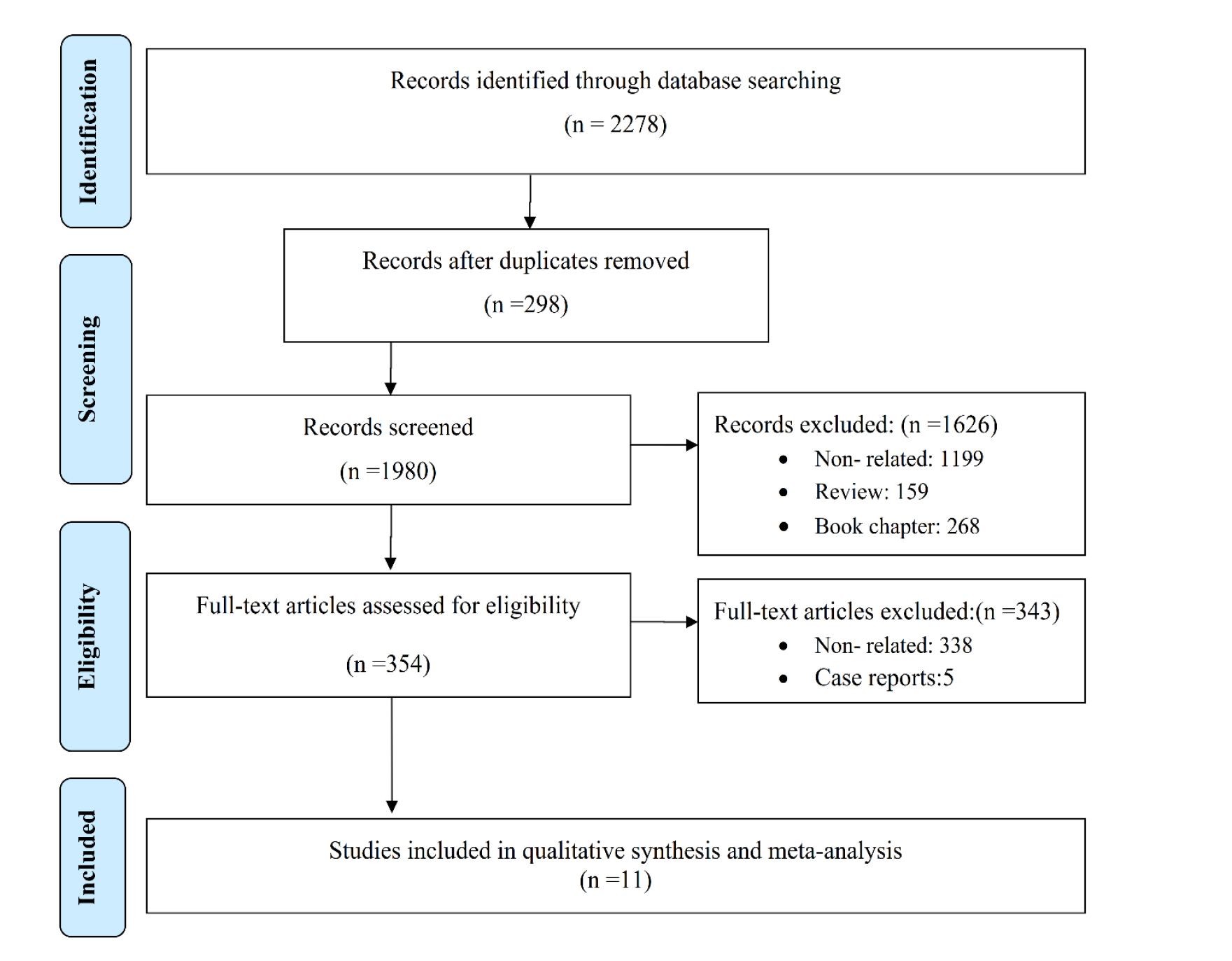
Figure 1. PRISMA flow diagram of the search strategy
The title and abstract screening were performed independently by two reviewers (J. J. and Z. T. H.). In case of conflict between the two reviewers, a third reviewer was included to solve the conflict. All initially searched articles were imported into the EndNote software (version 20, Clarivate Analytics, USA). The statistical analysis was performed using the StatsDirect (version 3, package StatsDirect Corp, Wirral, UK). The heterogeneity index for all studies was determined using the χ2-based weight of each study, and the horizontal lines drawn along the x-axis are 95% CI values. The funnel plot (Figure S1) and Egger's regression test were performed to check publication bias.
Results and Discussion
After the title and abstract screening, 354 relevant papers were obtained for full-paper screening. During the complete paper screening process, 343 papers were excluded as non-relevant or case reports. A data extraction Table 1 was used to report the following variables: author’s name, year of publication, study location, total otomycosis sample size, and Fusarium species (Table 1) [ 6 , 16 , 19 - 27 ].
| No. | Author/Publication Year/Reference | Location | Fusarium otomycosis no./Total sample no. (%) | Fusarium Spp. (No.) | Diagnostic method | Treatment | Study Period | Study population |
|---|---|---|---|---|---|---|---|---|
| 1 | Bassiouny et al. 1986 [ 19 ] | Egypt | 3/298 (1) | Fusarium Oxysporum (3) | Microscopy and culture | ND | ND | Otomycotic patients |
| 2 | Gugani et al.1989 [ 20 ] | Nigeria | 1/67 (1.49) | Fusarium spp. (1) | Microscopy and culture | ND | ND | Clinically suspected of otomycosis |
| 3 | Kombila et al.1989 [ 21 ] | Gabon | 1/83 (1.2) | Fusarium spp. (1) | Microscopy and culture | ND | ND | Otomycotic patients |
| 4 | Baveja et al.1993 [ 22 ] | India | 1/25 (4) | Fusarium spp. ( 1) | Microscopy and culture | Tolnaftate. | ND | Clinically suspected of otomycosis |
| 5 | Enweani et al.1997 [ 23 ] | Nigeria | 4/64 (6.25) | F. solani (4) | Microscopy and culture | ND | ND | Malnourished and healthy children |
| 6 | Jia et al. 2012 [ 16 ] | China | 1/108 (0.92) | F. solani (1) | Microscopy and culture | Topical Fluconazole | Sep 2009- Sep 2010 | Otomycotic patients |
| 7 | Kazemi et al. 2015 [ 24 ] | Iran | 2/129 (1.55) | Fusarium spp. (2) | Microscopy and culture | ND | 2009- 2011 | Clinically suspected of otomycosis |
| 8 | Kulal et al. 2017 [ 25 ] | India | 1/135 (0.74) | Fusarium spp. (1) | Microscopy and culture | ND | Nov 2008- Aug 2010 | Clinically suspected of otomycosis |
| 9 | Salari et al. 2017 [ 26 ] | Iran | 1/26 (3.84) | Fusarium spp. (1) | Microscopy, culture, and PCR | ND | Mar 2004- Mar 2014 | Suspected of Superficial and cutaneous fungal infections |
| 10 | Alarid-Coronel et al.2018 [ 6 ] | Mexico | 5/40 (12.5) | Fusarium spp. (5) | Microscopy and culture | Topical Antifungals | Aug 2010- Jan 2016 | Immunocompetent patients |
| 11 | Kiakojuri et al. 2019 [ 27 ] | Iran | 1/161 (0.62) | Fusarium spp. (1) | Microscopy and culture | Clotrimazole | ND | Clinically suspected of otomycosis |
| ND: not determined, PCR: polymerase chain reaction | ||||||||
Epidemiology of Fusarium otomycosis
A preliminary search of five databases yielded 2278 articles and 11 meta-analysed epidemiological studies for Fusarium otomycosis (Figure 1). The worldwide prevalence of otomycosis is estimated at approximately 1-4%. In the present review, the pooled prevalence of otomycosis due to Fusarium species is estimated at 2.3 (95% CI= 1.2-3.7) (Figure 2). The heterogeneity analysis of pooled data on all included studies revealed significantly high heterogeneity (Cochran Q= 18.5 (df= 10), p = 0.05) and (I2 =45.9% (95% CI= 0-71.6%). The highest prevalence was reported in Mexico (13%), and the lowest prevalence was related to Iran (0.62%). Among the Fusarium identified in otomycosis, the most common species of Fusarium were Fusarium spp. 13/21 (76.2%), followed by F. solani 5/21 (23.8%) and F. oxysporum 3/21 (14.3) (Table 1) [ 6 , 16 , 19 - 27 ].
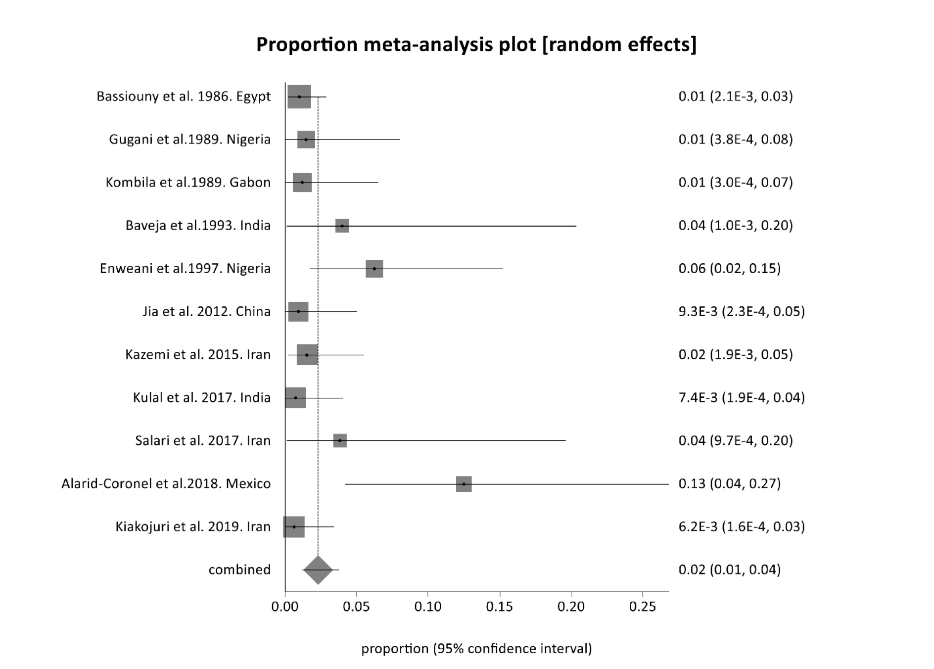
Figure 2. Forest plot for meta-analysis from 11 studies on Fusarium otomycosis
As the included otomycosis studies did not report gender and age distributions according to the causative fungal agent, the demographic analysis was conducted on the aggregate data. No consistent pattern was observed in terms of gender distribution, with the reported prevalence of otomycosis varying between male and female predominance across different studies [ 1 , 6 , 16 , 22 - 26 ]. This suggests that local environmental or occupational factors influence the condition rather than a fixed biological characteristic. The highest incidence rate of otomycosis was observed among young and middle-aged adults (21-40 years) [ 6 , 22 , 24 , 25 ]. While cases occur across all age groups, children and the elderly are less frequently affected. This demographic pattern may be linked to greater exposure to risk factors, such as swimming and earphone use, in younger populations.
Risk factors
Otomycosis refers to the superficial mycotic infection of the outer ear canal associated with various predisposing factors, such as alterations in ear pH and cerumen, swimming, external ear canal trauma or instrumentation, the use of hearing aid or ear prosthesis, bacterial infection, occupational exposure to a dry, dusty environment, inadequate hygiene, cleaning the external ear canal with matchsticks, and instilling oil and earwax in the external ear canal [ 4 , 5 , 28 - 30 ]. Two of the most critical factors for otomycosis are long-term moisture exposure and the use of topical antibiotics (fluoroquinolones) or steroid eardrops [ 4 , 14 , 31 ]. In addition, researchers acknowledge physiological conditions, such as pregnancy, diabetes mellitus, malignancy, and HIV infection, as significant predisposing factors [ 3 ].
Clinical manifestation
According to the results of the present meta-analysis, it is evident that Fusarium otomycosis is an uncommon disorder but clinically significant fungal infection [ 32 , 33 ]. The clinical manifestations are characterized by a broad spectrum of symptoms and signs not specific to Fusarium infection, resembling infections of the EAC caused by various agents. Individuals suffering from this condition typically experience symptoms, such as otalgia, otorrhea, and hearing impairment, which can progress if left untreated [ 34 ]. These initial symptoms can be followed by severe otalgia, frequently accompanied by pruritus, aural fullness, and discomfort. The presence of ear discharge, usually serous or serosanguinous, with a strong odour and debris build-up within the ear canal, can contribute to varying degrees of hearing loss, ranging from mild to severe infections [ 16 , 35 ]. Spread of the infection, especially if there is an extension to the tympanic membrane, can lead to sensorineural hearing loss if the middle ear is involved [ 14 ].
In complications, patients may experience less common symptoms, such as vertigo, tinnitus, and facial musculature weakness [ 36 ]. Physical examination in Fusarium otomycosis usually reveals erythematous and edematous ear canal skin, often accompanied by erosions and ulcerations [ 16 ]. In severe cases, mainly in immunocompromised patients, direct extension into adjacent structures, such as the temporomandibular joint or cranial nerves, may lead to further neurologic sequelae [ 37 ]. In differential diagnosis, it is essential to distinguish between this fungal infection and the other possible causes, such as acute otitis externa or chronic suppurative otitis media [ 2 ]. Moreover, early detection and identification of causative agents are crucial for the establishment of appropriate therapy, which can help prevent potential complications, including disease progression and associated morbidity [ 38 ].
Diagnostic approaches
Fusarium otomycosis is a relatively uncommon but increasingly recognized clinical entity that poses a diagnostic challenge due to its symptom similarity to other forms of ear infections, as well as the absence of standardized laboratory examinations. The initial steps in diagnosing approaches are gathering anamnestic data on symptoms and potential risk factors, followed by clinical examination-otoscopy. Knowledge of the ecological and epidemiological characteristics of the particular region is also essential in diagnosing this superficial fungal infection. During the clinical examination, healthcare professionals generally cannot differentiate Fusarium otomycosis from infection of a different etiology based solely on the observed symptoms and clinical signs [ 35 ]. Therefore, a laboratory diagnosis is necessary to manage this condition. If feasible, various diagnostic modalities are available, including conventional mycological techniques and molecular methods. Given that yeast and molds can be members of the microbiota naturally present on the skin of the EAC or represent fungal transient flora, the standard approach to address this dilemma is to perform multiple successive analyses using several samples, and in sporadic cases, confirmation can be achieved through histopathology [ 15 ].
Conventional mycological methods include microscopic examinations, which allow detecting fungal elements in samples but may not establish the specific etiological agents. On the other hand, cultivation represents the gold standard for isolation and identification of Fusarium species from clinical specimens. This culture-based mycological diagnosis includes inoculation of clinical specimens, such as ear discharge or tissue, onto appropriate fungal culture media (i.e., sabouraud dextrose agar and malt extract agar). The colonies that appear after 2-3 days of incubation at 25-30° care subsequently examined macroscopically and microscopically to identify the genus or species of fungi [ 2 ]. Molecular-based identification and matrix-assisted laser desorption/ionization-time of flight-mass spectrometry can be promising and robust tools to identify Fusarium species at the species level [ 39 ].
The DNA sequencing of the translation elongation factor 1α (TEF1α) gene has recently been developed to improve the sensitivity and specificity of the laboratory identification of Fusarium species [ 40 ]. Sequencing DNA and determining molecular targets for identification is an approach that could enable the design and establishment of rapid molecular testing for otomycosis [ 41 ]. Imaging studies, such as computed tomography or magnetic resonance imaging, may be used to determine the extent of the infection and to identify any underlying conditions that may be contributing to the infection [ 42 ]. It is important to note that the laboratory diagnosis of otomycosis due to Fusarium species can be challenging, and a combination of methods may be needed to confirm the diagnosis, since identification of Fusarium species is crucial for appropriate treatment.
Treatment options
Treatment of otomycosis is currently a big concern and challenge for otolaryngologists and physicians [ 7 ]. This is mainly due to the lack of established guidelines for diagnosis and treatment, and undefined therapy duration [ 15 , 43 , 44 ]. Considering their remarkably high prevalence, most infections of the external auditory canal are treated without reliance on laboratory-based evidence. Furthermore, inadequate laboratory practices, such as the omission of mycological examinations, as well as the fact that in some instances, mycological examination does not include the procedure for isolation of all potential causative fungi (contamination), make the treatment of otomycoses, including fusariosis, challenging. The issue becomes more serious when considering the potential resistance to currently available antifungal agents [ 7 ]. Dealing with infections caused by Fusarium species can be quite daunting, as clinically relevant Fusarium species are often found to be resistant to most antifungal agents, including azoles, echinocandins, and polyenes [ 18 ]. Additionally, it is worth emphasizing that Fusarium species exhibit intrinsic resistance to most currently available antifungal drug classes, such as azoles and echinocandins, even without prior exposure to these agents [ 18 , 45 ].
Findings of the present study and published data indicate that the successful treatment of otomycosis caused by Fusarium species is rare. Nevertheless, earlier studies have suggested that terbinafine could be a potential candidate for specific types of superficial infections caused by Fusarium species, supported by the results of a recent study conducted by Ting-Hua Yang, which demonstrated the non-toxic nature of this antimycotic to the inner ear end organs at a dosage of 0.4 mg [ 15 , 18 , 46 - 49 ]. Natamycin (5%), topical amphotericin B (0.5%), and 1% topical voriconazole for up to weeks are successfully employed to treat superficial Fusarium infections [ 50 , 51 ]. Echinocandins exhibit limited effectiveness against Fusarium species and are generally ineffective in treating infections caused by Fusarium species [ 18 ]. Combining antifungal agents has improved treatment efficacy in cases where mono-antifungal therapy proves ineffective [ 18 ]. Several in vitro or in vivo studies using an experimental murine model have explored the potential synergetic effect of combining various antifungal agents [ 52 - 54 ]. Terbinafine and voriconazole have exhibited a synergistic effect against different species of Fusarium; additionally, caspofungin, while demonstrating high minimum effective concentrations (MECs) against several Fusarium species when used alone, has shown a notably strong synergistic effect when combined with other antifungals [ 55 ]. In an experimental murine model, combination therapy of terbinafine and liposomal amphotericin B has shown promising results in treating Fusarium infections. Other antifungal agents, such as posaconazole or voriconazole in combination with amphotericin B, have displayed poor effectiveness against these species [ 56 , 57 ]. Al-Hatmi et al. found that a combination of natamycin and voriconazole demonstrated 70% in vitro synergistic interactions against a considerable portion of Fusarium isolates [ 58 ]. According to Spader et al., in vitro experiments revealed synergistic effects when amphotericin B was combined with rifampin, 5-flucytosine, caspofungin, and voriconazole [ 59 ]. Nosratabadi et al showed that luliconazole, lanoconazole, and efinaconazole had good activity against all Fusarium isolates, the minimum inhibitory concentration of luliconazole, lanoconazole and efinaconazole were in the ranges of 0.001–0.125, 0.001–0.5, and 0.064–4 μg/ml, respectively, and 272 of all isolates (96.4%) were inhibited in the concentration of ≤0.125 μg/ml of luliconazole and lanoconazole [ 60 ]. Nosratabadi et al. investigated the in vitro antifungal susceptibility pattern of miltefosine against a collection of azole and echinocandin-resistant Fusarium strains. Their results revealed that amphotericin B (0.8µg/mL) had the lowest geometric mean MICs/MECs values, followed by miltefosine (1.44µg/mL), voriconazole (2.15µg/mL), caspofungin (7.23µg/mL), and itraconazole (14.19µg/mL). Therefore, after amphotericin B, miltefosine has shown superior efficacy against Fusarium isolates, compared to azoles and echinocandins. This suggests its potential as a novel treatment for Fusarium infections and warrants further investigation through in vivo efficacy studies [ 61 ]. More clinical studies are needed to examine the most effective combination for the treatment of Fusarium infections, and additional research is recommended to address this issue [ 18 ].
Conclusion
Otitis externa is an infection of the ear canal caused by bacteria and fungi (otomycosis), which can cause pain due to inflammation. If otitis externa does not respond to treatment and involves the bony structures, it can progress to malignant otitis externa. This infection can spread from the outer ear to nearby tissues and the temporal bone through the fissure of Santorini. Findings of the present study, together with published data, indicate that otomycosis caused by Fusarium species is an emerging clinical entity that warrants attention. Awareness among physicians and the use of appropriate diagnostic techniques are essential for the timely identification and effective management of infections. Considering the potential complications, such as direct extension into joint or cranial nerves, as well as the resistance of Fusarium species to regular antifungal agents with a different mechanism of action, it is crucial to include otomycosis caused by non-dermatophyte fungi in the differential diagnosis, followed by effective treatment and monitoring.
Acknowledgments
Sincere appreciation and thanks are extended to the authors for their support and contributions.
Conflicts of interest
No conflict of interest is declared.
Authors’ contributions
Conceptualization: I. H. and M. A.; Literature search: Z. T. R., F. K., J. J., M. GH., B. R., M. GH., and SH. KH.; Data collection: J. J. and M. N.; Data analysis: J. J., R. SH., and F. K.; Data interpretation: I. H., M. A., and S. O.; Resources: J. J., R. SH., M. GH.; Writing-original draft preparation: I. H., B. R., Z. F. and F. K.; Writing-review and editing: H. B., A. AL. H., M. H., and S. O.; Statistical analysis: GH. SH.; Supervision: I. H. and M. A. All authors have read and agreed to the published version of the manuscript.
Financial disclosure
This research was funded by the Mazandaran University of Medical Sciences [grant number 14961].
Ethics approval
The current study was approved by the Ethics Committee of the Mazandaran University of Medical Sciences (IR.MAZUMS.REC.1401.469).
References
- Kiakojuri K, Mahdavi Omran S, Roodgari S, Taghizadeh Armaki M, Hedayati MT, Shokohi T, et al. Molecular identification and antifungal susceptibility of yeasts and molds isolated from patients with otomycosis. Mycopathologia. 2021; 186(2):245-57.
- Gharaghani M, Seifi Z, Zarei Mahmoudabadi A. Otomycosis in iran: a review. Mycopathologia. 2015; 179(5-6):415-24.
- Fasunla J, Ibekwe T, Onakoya P. Otomycosis in western Nigeria. Mycoses. 2008; 51(1):67-70.
- Munguia R, Daniel SJ. Ototopical antifungals and otomycosis: a review. Int J Pediatr Otorhinolaryngol. 2008; 72(4):453-9.
- Barati B, Okhovvat SA, Goljanian A, Omrani MR. Otomycosis in central iran: a clinical and mycological study. Iran Red Crescent Med J. 2011; 13(12):873-6.
- Alarid-Coronel J, Celis-Aguilar E, Escobar-Aispuro L, Muñoz-Estrada V. Otomycosis in immunocompetent patients: Clinical and mycological features. Our experience with 40 cases. Clin Otolaryngol 2018; 43:373-7.
- Sangare I, Amona FM, Ouedraogo RW, Zida A, Ouedraogo MS. Otomycosis in Africa: Epidemiology, diagnosis and treatment. J Mycol Med. 2021; 31(2):101115.
- Abastabar M, Haghani I, Ahangarkani F, Rezai MS, Taghizadeh Armaki M, Roodgari S, et al. Candida auris otomycosis in Iran and review of recent literature. Mycoses. 2019; 62(2):101-5.
- Armaki MT, Omran SM, Kiakojuri K, Khojasteh S, Jafarzadeh J, Tavakoli M, et al. First fluconazole-resistant Candida auris isolated from fungal otitis in Iran. Curr Med Mycol. 2021; 7(1)
- Ahmadi B, Naeimi B, Ahmadipour MJ, Morovati H, de Groot T, Spruijtenburg B, et al. An autochthonous susceptible Candida auris clade I otomycosis case in Iran. J Fungi. 2023; 9(11):1101.
- Chowdhary A, Prakash A, Sharma C, Kordalewska M, Kumar A, Sarma S, et al. A multicentre study of antifungal susceptibility patterns among 350 Candida auris isolates (2009–17) in India: role of the ERG11 and FKS1 genes in azole and echinocandin resistance. J Antimicrob Chemother. 2018; 73(4):891-9.
- Ali K, Hamed MA, Hassan H, Esmail A, Sheneef A. Identification of fungal pathogens in otomycosis and their drug sensitivity: our experience. Int Archiv Otorhinolaryngol. 2018; 22(04):400-3.
- Ho T, Vrabec JT, Yoo D, Coker NJ. Otomycosis: clinical features and treatment implications. Otolaryngol Head Neck Surg. 2006; 135(5):787-91.
- Vennewald I, Klemm E. Otomycosis: Diagnosis and treatment. Clin Dermatol. 2010; 28(2):202-11.
- Bojanović M, Stalević M, Arsić-Arsenijević V, Ignjatović A, Ranđelović M, Golubović M, et al. Etiology, predisposing factors, clinical features and diagnostic procedure of otomycosis: a literature review. J Fungi (Basel). 2023; 9(6):662.
- Jia X, Liang Q, Chi F, Cao W. Otomycosis in Shanghai: aetiology, clinical features and therapy. Mycoses. 2012; 55(5):404-9.
- Saki N, Rafiei A, Nikakhlagh S, Amirrajab N, Saki S. Prevalence of otomycosis in Khouzestan Province, south-west Iran. J Laryngol Otol. 2013; 127(1):25-7.
- Al-Hatmi AM, Meis JF, de Hoog GS. Fusarium: Molecular diversity and intrinsic drug resistance. PLoS Pathog. 2016; 12(4):e1005464.
- Bassiouny A, Kamel T, Moawad M, Hindawy D. Broad spectrum antifungal agents in otomycosis. J Laryngol Otol. 1986; 100(8):867-73.
- Gugnani H, Okafor B, Nzelibe F, Njoku‐Obi A. Etiological agents of otomycosis in nigeria das erregerspektrum von otomykosen in Nigeria. Mycoses. 1989; 32(5):224-9.
- Kombila M, Gomez de Diaz M, De Bievre C, Crepet G, Debrie J, Belembaogo E, et al. Fungal otitis in Libreville. Study of 83 cases. Bulletin de la Société de pathologie exotique et de ses filiales. 1989; 82(2):201-7.
- Baveja C, Dhingra P, Natarajan R. Mycological profile in otomycosis and its response to an antifungal agent. Indian J Otolaryngol Head Neck Surg. 1993; 45:224-5.
- Enweani I, Igumbor H. Prevalence of otomycosis in malnourished children in Edo State, Nigeria. Mycopathologia. 1997; 140(2):85-7.
- Kazemi A, Majidinia M, Jaafari A, Mousavi SAA, Mahmoudabadi AZ, Alikhah H. Etiologic agents of otomycosis in the North-Western area of Iran. Jundishapur J Microbiol. 2015; 8(9):e21776.
- Kulal B, Bhat KS, Meundi M, Kotigadde S. A microbiological study of otomycosis. Indian J Microbiol Res. 2017; 4(1):118-25.
- Salari S, Mousavi SAA, Hadizadeh S, Izadi A. Epidemiology of dermatomycoses in Kerman province, southeast of Iran: a 10-years retrospective study (2004–2014). Microb Pathog. 2017; 110:561-7.
- Kiakojuri K, Rajabnia R, Mahdavi Omran S, Pournajaf A, Karami M, Taghizadeh Armaki M. Role of clotrimazole in prevention of recurrent otomycosis. BioMed Res Int. 2019; 2019:5269535.
- Dundar R, İynen İ. Single dose topical application of clotrimazole for the treatment of otomycosis: is this enough? J Audiol Otol. 2019; 23(1):15.
- Prasad SC, Kotigadde S, Shekhar M, Thada ND, Prabhu P, T DS, et al. Primary otomycosis in the Indian subcontinent: predisposing factors, microbiology, and classification. Int J Microbiol. 2014; 2014:636493.
- Khojasteh S, Jafarzdeh J, Hosseini SA, Haghani I, Turki H, Gharehbolagh SA, et al. Candida auris and COVID-19: A health threatening combination. Cur Med Mycol. 2022; 8(3):44.
- Nowrozi H, Arabi FD, Mehraban HG, Tavakoli A, Ghooshchi G. Mycological and clinical study of otomycosis in Tehran, Iran. Bull Environ Pharmacol Life Sci. 2014; 3(2):29-31.
- Nnadi NE, Carter DA. Climate change and the emergence of fungal pathogens. PLoS Pathog. 2021; 17(4):e1009503.
- Khattab AA, Swelim M, Abdelsater MA, Ibrahim G, Abdelrahman A. Fungi associated within human otomycosis. Egyp Acad J Biol Sci G Microbiol. 2022; 14(1):113-23.
- Cheraghsahar S, Kazemi S, Birjandi M, Yarahmadi M, Mahmoudi S, Mohammadi R, et al. Otomycosis in western Iran: clinical and mycological aspects. Arch Clin Infect Dis. 2017; 12(2)
- Roohi B, Nemati S, Alipour A, Faeli L, Mayahi S, Haghani I, et al. Otomycosis: the foremost aetiological agent causing otitis externa and the antifungal susceptibility pattern in North-Western Iran. Mycoses. 2023; 66(2):87-97.
- Munagala K, Mood N, Baddam GSC. Efficacy of 7.5% povidone-iodine and 1% clotrimazole in the treatment of otomycosis-A prospective randomized study. Natl J Physiol Pharm Pharmacol. 2022; 12(8):1142-6.
- Douglas AP, Chen SC, Slavin MA. Emerging infections caused by non-Aspergillus filamentous fungi. Clin Microbiol Infect. 2016; 22(8):670-80.
- Gu X, Cheng X, Zhang J, She W. Identification of the fungal community in otomycosis by internal transcribed spacer sequencing. Front Microbiol. 2022; 13:820423.
- Haghani I, Shams-Ghahfarokhi M, Dalimi Asl A, Shokohi T, Hedayati MT. Prevalence, genetic diversity and antifungal susceptibility profiles of F. fujikuroi, F. solani and Fusarium incarnatum-equiseti species complexes from onychomycosis in north of Iran. Mycoses. 2022; 65(11):1030-9.
- Dallé da Rosa P, Aquino V, Fuentefria AM, Goldani LZ. Diversity of Fusarium species causing invasive and disseminated infections. J Med Mycol. 2021; 31(3):101137.
- Hagiwara S, Tamura T, Satoh K, Kamewada H, Nakano M, Shinden S, et al. The molecular identification and antifungal susceptibilities of aspergillus species causing otomycosis in Tochigi, Japan. Mycopathologia. 2019; 184(1):13-21.
- Lawal IO, Mokoala KM, Kgatle MM, Dierckx RA, Glaudemans AW, Sathekge MM, et al. Radionuclide imaging of invasive fungal disease in immunocompromised hosts. Diagnostics. 2021; 11(11):2057.
- Bojanovic M, Ignjatovic A, Stalevic M, Arsic-Arsenijevic V, Randelovic M, Gerginic V, et al. Clinical presentations, cluster analysis and laboratory-based investigation of aspergillus otomycosis-a single center experience. J Fungi (Basel). 2022; 8(3):315.
- Chappe M, Vrignaud S, de Gentile L, Legrand G, Lagarce F, Le Govic Y. Successful treatment of a recurrent Aspergillus niger otomycosis with local application of voriconazole. J Mycol Med. 2018; 28(2):396-8.
- Al-Hatmi AM, van Diepeningen AD, Curfs-Breuker I, de Hoog GS, Meis JF. Specific antifungal susceptibility profiles of opportunists in the Fusarium fujikuroi complex. J Antimicrob Chemother. 2015; 70(4):1068-71.
- Ghannoum M, Isham N. Fungal nail infections (onychomycosis): a never-ending story? PLoS Pathog. 2014; 10(6):e1004105.
- Yang TH, Young YH. Eradicating otomycosis with terbinafine solution: basic and clinical investigation. Audiol Neurootol. 2019; 24(4):183-190.
- Xu S, Li J, Ding L, Gao K, Xie F, Han J, et al. Efficacy and safety of terbinafine hydrochloride spray and 3% boric acid alcohol ear drops in otomycosis. Acta Otolaryngol. 2020; 140(4):302-6.
- Morteza-Semnani K, Saeedi M, Akbari J, Hedayati S, Hashemi SMH, Rahimnia SM, et al. Green formulation, characterization, antifungal and biological safety evaluation of terbinafine HCl niosomes and niosomal gels manufactured by eco-friendly green method. J Biomater Sci Polym Ed. 2022; 33(18):2325-52.
- Sharma S, Das S, Virdi A, Fernandes M, Sahu SK, Kumar Koday N, Ali MH, Garg P, et al. Re-appraisal of topical 1% voriconazole and 5% natamycin in the treatment of fungal keratitis in a randomised trial. Br J Ophthalmol. 2015; 99(9):1190-5.
- Edwar L, Janna YM, Rozaliyani A, Louisa M. Therapeutic response time of topical voriconazole 1% and intrastromal voriconazole 0.05% versus topical natamycin 5% monotherapy in Fusarium keratitis in rabbit. Mycoses. 2020; 63(10):1128-32.
- Ruíz-Cendoya M, Mariné M, Guarro J. Combined therapy in treatment of murine infection by Fusarium solani. J Antimicrob Chemother. 2008; 62(3):543-6.
- Ruíz-Cendoya M, Mariné M, Rodríguez MM, Guarro J. Interactions between triazoles and amphotericin B in treatment of disseminated murine infection by Fusarium oxysporum. Antimicrob Agents Chemother. 2009; 53(4):1705-8.
- Bidaud A-L, Schwarz P, Herbreteau G, Dannaoui E. Techniques for the assessment of in vitro and in vivo antifungal combinations. J Fungi. 2021; 7(2):113.
- Guarro J. Fusariosis, a complex infection caused by a high diversity of fungal species refractory to treatment. Eur J Clin Microbiol Infect Dis. 2013; 32(12):1491-1500.
- Ruiz-Cendoya M, Pastor FJ, Capilla J, Guarro J. Treatment of murine Fusarium verticillioides infection with liposomal amphotericin B plus terbinafine. Int J Antimicrob Agents. 2011; 37(1):58-61.
- Ruíz-Cendoya M, Pastor J, Guarro J. Combined therapy against murine-disseminated infection by Fusarium verticillioides. Mycopathologia. 2011; 171:171-5.
- Al-Hatmi AM, Meletiadis J, Curfs-Breuker I, Bonifaz A, Meis JF, De Hoog G. In vitro combinations of natamycin with voriconazole, itraconazole and micafungin against clinical Fusarium strains causing keratitis. J Antimicrob Chemother. 2016; 71(4):953-5.
- Spader TB, Venturini TP, Rossato L, Denardi LB, Cavalheiro PB, Botton SA, et al. Synergysm of voriconazole or itraconazole with other antifungal agents against species of Fusarium. Rev Iberoam Micol. 2013; 30(3):200-4.
- Nosratabadi M, Faeli L, Haghani I, Mohammadi R, Khodavaisy S, Kachuei R, et al. In vitro antifungal susceptibility profile of Iranian Fusarium isolates: Emphasising on the potent inhibitory effect of efinaconazole compared to other drugs. Mycoses. 2023; 66(3):258-75.
- Nosratabadi M, Akhtari J, Faeli L, Haghani I, Aghili SR, Shokohi T, et al. In vitro antifungal susceptibility profile of Miltefosine against a collection of azole and echinocandins resistant Fusarium strains. J Fungi. 2022; 8(7):709.


